 Movie
Movie Controller

[English] 日本語
 Yorodumi
- PDB-6zpk: Crystal structure of the unconventional kinetochore protein Trypa... -
+ Open data

- Basic information
| Entry | Database: PDB / ID: 6zpk | ||||||
|---|---|---|---|---|---|---|---|
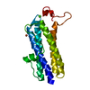
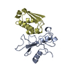
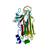
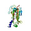
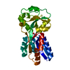
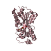
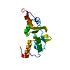
| Title | Crystal structure of the unconventional kinetochore protein Trypanosoma brucei KKT4 BRCT domain | ||||||
 Components Components | Trypanosoma brucei KKT4 463-645 | ||||||
 Keywords Keywords | CELL CYCLE / kinetochore / kinetoplastid / KKT4 / BRCT | ||||||
| Function / homology |  Function and homology information Function and homology informationspindle microtubule / chromosome segregation / kinetochore / microtubule binding / nucleus Similarity search - Function | ||||||
| Biological species |  | ||||||
| Method |  X-RAY DIFFRACTION / SYNCHROTRON / AB INITIO PHASING / Resolution: 1.57 Å X-RAY DIFFRACTION / SYNCHROTRON / AB INITIO PHASING / Resolution: 1.57 Å | ||||||
 Authors Authors | Ludzia, P. / Lowe, E.D. / Marciano, G. / Mohammed, S. / Redfield, C. / Akiyoshi, B. | ||||||
| Funding support |  United Kingdom, 1items United Kingdom, 1items
| ||||||
 Citation Citation | Journal: Structure / Year: 2021 Title: Structural characterization of KKT4, an unconventional microtubule-binding kinetochore protein. Authors: Ludzia, P. / Lowe, E.D. / Marciano, G. / Mohammed, S. / Redfield, C. / Akiyoshi, B. #1: Journal: Biorxiv / Year: 2020Title: Structural characterisation of KKT4, an unconventional microtubule-binding kinetochore protein Authors: Ludzia, P. / Lowe, E. / Marciano, G. / Mohammed, S. / Redfield, C. / Akiyoshi, B. | ||||||
| History |
|
- Structure visualization
| Structure viewer | Molecule: MolmilJmol/JSmol |
|---|
- Downloads & links
-Download
| PDBx/mmCIF format | 6zpk.cif.gz | 87.6 KB | Display | PDBx/mmCIF format |
|---|---|---|---|---|
| PDB format | pdb6zpk.ent.gz | 64.8 KB | Display | PDB format |
| PDBx/mmJSON format | 6zpk.json.gz | Tree view | PDBx/mmJSON format | |
| Others |  Other downloads Other downloads |
-Validation report
| Arichive directory | https://data.pdbj.org/pub/pdb/validation_reports/zp/6zpkftp://data.pdbj.org/pub/pdb/validation_reports/zp/6zpk | HTTPS FTP |
|---|
-Related structure data


-Links
 PDBj
PDBj

- Assembly
| Deposited unit | 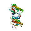
| ||||||||||
|---|---|---|---|---|---|---|---|---|---|---|---|
| 1 |
| ||||||||||
| Unit cell |
|
-Components
| #1: Protein | Mass: 20130.969 Da / Num. of mol.: 1 Source method: isolated from a genetically manipulated source Details: Highlighted residues are not visible in the electron density. Trypanosoma brucei KKT4 463-645 Source: (gene. exp.) Strain: 927/4 GUTat10.1 / Gene: Tb927.8.3680 / Plasmid: pNIC28-Bsa4 / Production host:  |
|---|---|
| #2: Chemical | ChemComp-GOL /   Mass: 92.094 Da / Num. of mol.: 1 / Source method: obtained synthetically / Formula: C3H8O3 Mass: 92.094 Da / Num. of mol.: 1 / Source method: obtained synthetically / Formula: C3H8O3 |
| #3: Chemical | ChemComp-SO4 /   Mass: 96.063 Da / Num. of mol.: 1 / Source method: obtained synthetically / Formula: SO4 Mass: 96.063 Da / Num. of mol.: 1 / Source method: obtained synthetically / Formula: SO4 |
| #4: Water | ChemComp-HOH /  Mass: 18.015 Da / Num. of mol.: 205 / Source method: isolated from a natural source / Formula: H2O Mass: 18.015 Da / Num. of mol.: 205 / Source method: isolated from a natural source / Formula: H2O |
| Has ligand of interest | N |
-Experimental details
-Experiment
| Experiment | Method: X-RAY DIFFRACTION / Number of used crystals: 1 |
|---|
- Sample preparation
| Crystal | Density Matthews: 2.41 Å3/Da / Density % sol: 48.87 % |
|---|---|
| Crystal grow | Temperature: 277 K / Method: vapor diffusion, sitting drop / pH: 5.5 Details: HEPES, sodium chloride, bis-Tris, ammonium sulphate, TCEP |
-Data collection
| Diffraction | Mean temperature: 100 K / Serial crystal experiment: N |
|---|---|
| Diffraction source | Source: SYNCHROTRON / Site: Diamond / Beamline: I24 / Wavelength: 0.9686 Å |
| Detector | Type: DECTRIS PILATUS3 6M / Detector: PIXEL / Date: Feb 12, 2018 |
| Radiation | Protocol: SINGLE WAVELENGTH / Monochromatic (M) / Laue (L): M / Scattering type: x-ray |
| Radiation wavelength | Wavelength: 0.9686 Å / Relative weight: 1 |
| Reflection | Resolution: 1.56956→45.6013 Å / Num. obs: 27084 / % possible obs: 97.3 % / Redundancy: 10 % / Biso Wilson estimate: 15.7 Å2 / CC1/2: 0.997 / Net I/σ(I): 14 |
| Reflection shell | Resolution: 1.57→1.6 Å / Rmerge(I) obs: 0.549 / Num. unique obs: 1025 / CC1/2: 0.496 / Rpim(I) all: 0.319 / Rrim(I) all: 0.641 |
- Processing
| Software |
| |||||||||||||||||||||
|---|---|---|---|---|---|---|---|---|---|---|---|---|---|---|---|---|---|---|---|---|---|---|
| Refinement | Method to determine structure: AB INITIO PHASING / Resolution: 1.57→45.6 Å / SU ML: 0.1434 / Cross valid method: FREE R-VALUE / σ(F): 1.34 / Phase error: 20.1667 / Stereochemistry target values: GeoStd + Monomer Library
| |||||||||||||||||||||
| Solvent computation | Shrinkage radii: 0.9 Å / VDW probe radii: 1.11 Å / Solvent model: FLAT BULK SOLVENT MODEL | |||||||||||||||||||||
| Displacement parameters | Biso mean: 23.04 Å2 | |||||||||||||||||||||
| Refinement step | Cycle: LAST / Resolution: 1.57→45.6 Å
|
