 Movie
Movie Controller

[English] 日本語
 Yorodumi
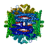
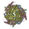
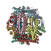
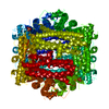
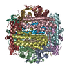
- PDB-6tgt: The Calcium soaked crystal structure of the DPS2 from DEINOCOCCUS... -
+ Open data

- Basic information
| Entry | Database: PDB / ID: 6tgt | ||||||
|---|---|---|---|---|---|---|---|
| Title | The Calcium soaked crystal structure of the DPS2 from DEINOCOCCUS RADIODURANS to 2.16A resolution (Soaked in CaCl2 [5mM] for 20 min). | ||||||
 Components Components | DNA protection during starvation protein 2 | ||||||
 Keywords Keywords | DNA BINDING PROTEIN / Iron biomineralisation / iron storage / iron detoxification / DNA binding / Calcium binding / metalloprotein. | ||||||
| Function / homology |  Function and homology information Function and homology informationDnaA-Dps complex / Oxidoreductases; Oxidizing metal ions / ferric iron binding / negative regulation of DNA-templated DNA replication initiation / intracellular iron ion homeostasis / oxidoreductase activity / DNA binding / cytoplasm Similarity search - Function | ||||||
| Biological species |  Deinococcus radiodurans (radioresistant) Deinococcus radiodurans (radioresistant) | ||||||
| Method |  X-RAY DIFFRACTION / SYNCHROTRON / MOLECULAR REPLACEMENT / Resolution: 2.155 Å X-RAY DIFFRACTION / SYNCHROTRON / MOLECULAR REPLACEMENT / Resolution: 2.155 Å | ||||||
 Authors Authors | Cuypers, M.G. / Romao, C.V. / Mitchell, E.P. / McSweeney, S. | ||||||
 Citation Citation | Journal: To Be Published Title: The Calcium soaked crystal structure of the DPS2 from DEINOCOCCUS RADIODURANS to 2.16A resolution (Soaked in CaCl2 [5mM] for 20 min). Authors: Cuypers, M.G. / McSweeney, S. / Romao, C.V. / Mitchell, E.P. | ||||||
| History |
|
- Structure visualization
| Structure viewer | Molecule: MolmilJmol/JSmol |
|---|
- Downloads & links
-Download
| PDBx/mmCIF format | 6tgt.cif.gz | 51.9 KB | Display | PDBx/mmCIF format |
|---|---|---|---|---|
| PDB format | pdb6tgt.ent.gz | 35.5 KB | Display | PDB format |
| PDBx/mmJSON format | 6tgt.json.gz | Tree view | PDBx/mmJSON format | |
| Others |  Other downloads Other downloads |
-Validation report
| Arichive directory | https://data.pdbj.org/pub/pdb/validation_reports/tg/6tgtftp://data.pdbj.org/pub/pdb/validation_reports/tg/6tgt | HTTPS FTP |
|---|
-Related structure data
| Related structure data |  2c2jS S: Starting model for refinement |
|---|---|
| Similar structure data | |
| Other databases |
|


-Links
 PDBj
PDBj


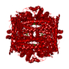
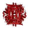
- Assembly
| Deposited unit | 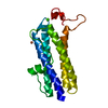
| ||||||||||||||||||
|---|---|---|---|---|---|---|---|---|---|---|---|---|---|---|---|---|---|---|---|
| 1 | x 12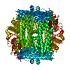
| ||||||||||||||||||
| Unit cell |
| ||||||||||||||||||
| Components on special symmetry positions |
|
-Components
| #1: Protein | Mass: 23075.469 Da / Num. of mol.: 1 Source method: isolated from a genetically manipulated source Source: (gene. exp.) Deinococcus radiodurans (strain ATCC 13939 / DSM 20539 / JCM 16871 / LMG 4051 / NBRC 15346 / NCIMB 9279 / R1 / VKM B-1422) (radioresistant)Strain: ATCC 13939 / DSM 20539 / JCM 16871 / LMG 4051 / NBRC 15346 / NCIMB 9279 / R1 / VKM B-1422 Gene: dps2, dps-2, DR_B0092 / Plasmid: PDEST14 / Production host: References: UniProt: Q9RZN1, Oxidoreductases; Oxidizing metal ions | ||||
|---|---|---|---|---|---|
| #2: Chemical | ChemComp-FE /   Mass: 55.845 Da / Num. of mol.: 1 / Source method: obtained synthetically / Formula: Fe / Feature type: SUBJECT OF INVESTIGATION Mass: 55.845 Da / Num. of mol.: 1 / Source method: obtained synthetically / Formula: Fe / Feature type: SUBJECT OF INVESTIGATION | ||||
| #3: Chemical |   Mass: 40.078 Da / Num. of mol.: 3 / Source method: obtained synthetically / Formula: Ca / Feature type: SUBJECT OF INVESTIGATION Mass: 40.078 Da / Num. of mol.: 3 / Source method: obtained synthetically / Formula: Ca / Feature type: SUBJECT OF INVESTIGATION#4: Water | ChemComp-HOH / |  Mass: 18.015 Da / Num. of mol.: 115 / Source method: isolated from a natural source / Formula: H2O Mass: 18.015 Da / Num. of mol.: 115 / Source method: isolated from a natural source / Formula: H2OHas ligand of interest | Y | |
-Experimental details
-Experiment
| Experiment | Method: X-RAY DIFFRACTION / Number of used crystals: 1 |
|---|
- Sample preparation
| Crystal | Density Matthews: 2.47 Å3/Da / Density % sol: 50.3 % / Description: cube |
|---|---|
| Crystal grow | Temperature: 293 K / Method: vapor diffusion, hanging drop / pH: 7.5 Details: 0.1 M KCl, 0.01 M MgCl2, 0.05 M HEPES pH 7.0, 5% v/v PEG 400 (Hampton Natrix crystal screen # 35) Temp details: regulated temperature room |
-Data collection
| Diffraction | Mean temperature: 100 K / Ambient temp details: cryostream N2 / Serial crystal experiment: N | ||||||||||||||||||||||||||||||
|---|---|---|---|---|---|---|---|---|---|---|---|---|---|---|---|---|---|---|---|---|---|---|---|---|---|---|---|---|---|---|---|
| Diffraction source | Source: SYNCHROTRON / Site: ESRF  / Beamline: ID29 / Wavelength: 0.9537 Å / Beamline: ID29 / Wavelength: 0.9537 Å | ||||||||||||||||||||||||||||||
| Detector | Type: DECTRIS PILATUS 6M / Detector: PIXEL / Date: Dec 7, 2005 | ||||||||||||||||||||||||||||||
| Radiation | Protocol: SINGLE WAVELENGTH / Monochromatic (M) / Laue (L): M / Scattering type: x-ray | ||||||||||||||||||||||||||||||
| Radiation wavelength | Wavelength: 0.9537 Å / Relative weight: 1 | ||||||||||||||||||||||||||||||
| Reflection | Resolution: 2.155→51 Å / Num. obs: 12682 / % possible obs: 100 % / Redundancy: 10.6 % / Biso Wilson estimate: 28.4 Å2 / CC1/2: 0.998 / Rmerge(I) obs: 0.127 / Rpim(I) all: 0.041 / Rrim(I) all: 0.133 / Χ2: 0.92 / Net I/σ(I): 17.2 | ||||||||||||||||||||||||||||||
| Reflection shell | Diffraction-ID: 1
|
- Processing
| Software |
| ||||||||||||||||||||||||
|---|---|---|---|---|---|---|---|---|---|---|---|---|---|---|---|---|---|---|---|---|---|---|---|---|---|
| Refinement | Method to determine structure: MOLECULAR REPLACEMENT Starting model: 2c2j Resolution: 2.155→44.21 Å / Cor.coef. Fo:Fc: 0.944 / Cor.coef. Fo:Fc free: 0.908 / SU B: 0.004 / SU ML: 0 / Cross valid method: THROUGHOUT / σ(F): 0 / ESU R: 0.157 / ESU R Free: 0.177 Details: HYDROGENS HAVE BEEN ADDED IN THE RIDING POSITIONS U VALUES : REFINED INDIVIDUALLY
| ||||||||||||||||||||||||
| Solvent computation | Ion probe radii: 0.8 Å / Shrinkage radii: 0.8 Å / VDW probe radii: 1.2 Å | ||||||||||||||||||||||||
| Displacement parameters | Biso max: 86.16 Å2 / Biso mean: 36.114 Å2 / Biso min: 17.55 Å2
| ||||||||||||||||||||||||
| Refinement step | Cycle: final / Resolution: 2.155→44.21 Å
| ||||||||||||||||||||||||
| LS refinement shell | Resolution: 2.157→2.213 Å / Rfactor Rfree error: 0
|
