 Movie
Movie Controller

[English] 日本語
 Yorodumi
- PDB-6tb5: The crystal structure of the DPS2 from DEINOCOCCUS RADIODURANS to... -
+ Open data

- Basic information
| Entry | Database: PDB / ID: 6tb5 | ||||||
|---|---|---|---|---|---|---|---|
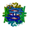
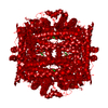
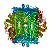
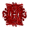
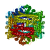
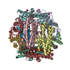
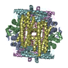
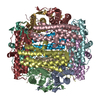
| Title | The crystal structure of the DPS2 from DEINOCOCCUS RADIODURANS to 1.83A resolution (sequentially soaked in CaCl2 [5mM] for 20 min, then in Ammonium iron(II) sulfate [10mM] for 2h). | ||||||
 Components Components | DNA protection during starvation protein 2 | ||||||
 Keywords Keywords | DNA BINDING PROTEIN / Iron biomineralisation / iron storage / iron detoxification / DNA binding / Calcium binding. | ||||||
| Function / homology |  Function and homology information Function and homology informationDnaA-Dps complex / Oxidoreductases; Oxidizing metal ions / ferric iron binding / negative regulation of DNA-templated DNA replication initiation / intracellular iron ion homeostasis / oxidoreductase activity / DNA binding / cytoplasm Similarity search - Function | ||||||
| Biological species |  Deinococcus radiodurans (radioresistant) Deinococcus radiodurans (radioresistant) | ||||||
| Method |  X-RAY DIFFRACTION / SYNCHROTRON / MOLECULAR REPLACEMENT / Resolution: 1.83 Å X-RAY DIFFRACTION / SYNCHROTRON / MOLECULAR REPLACEMENT / Resolution: 1.83 Å | ||||||
 Authors Authors | Cuypers, M.G. / McSweeney, S. / Romao, C.V. / Mitchell, E.P. | ||||||
 Citation Citation | Journal: To Be Published Title: The crystal structure of the DPS2 from DEINOCOCCUS RADIODURANS to 1.83A resolution (sequentially soaked in CaCl2 [5mM] for 20 min, then in Ammonium iron(II) sulfate [10mM] for 2h). Authors: Cuypers, M.G. / McSweeney, S. / Romao, C.V. / Mitchell, E.P. | ||||||
| History |
|
- Structure visualization
| Structure viewer | Molecule: MolmilJmol/JSmol |
|---|
- Downloads & links
-Download
| PDBx/mmCIF format | 6tb5.cif.gz | 54.3 KB | Display | PDBx/mmCIF format |
|---|---|---|---|---|
| PDB format | pdb6tb5.ent.gz | 37 KB | Display | PDB format |
| PDBx/mmJSON format | 6tb5.json.gz | Tree view | PDBx/mmJSON format | |
| Others |  Other downloads Other downloads |
-Validation report
| Arichive directory | https://data.pdbj.org/pub/pdb/validation_reports/tb/6tb5ftp://data.pdbj.org/pub/pdb/validation_reports/tb/6tb5 | HTTPS FTP |
|---|
-Related structure data
| Related structure data |  2c6rS S: Starting model for refinement |
|---|---|
| Similar structure data |

-Links
 PDBj
PDBj


- Assembly
| Deposited unit | 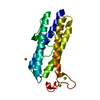
| |||||||||||||||||||||
|---|---|---|---|---|---|---|---|---|---|---|---|---|---|---|---|---|---|---|---|---|---|---|
| 1 | x 12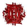
| |||||||||||||||||||||
| Unit cell |
| |||||||||||||||||||||
| Components on special symmetry positions |
|
-Components
| #1: Protein | Mass: 23289.715 Da / Num. of mol.: 1 Source method: isolated from a genetically manipulated source Source: (gene. exp.) Deinococcus radiodurans (radioresistant)Gene: dps2, dps-2, DR_B0092 / Plasmid: PDEST14 / Production host: References: UniProt: Q9RZN1, Oxidoreductases; Oxidizing metal ions | ||||||
|---|---|---|---|---|---|---|---|
| #2: Chemical |   Mass: 40.078 Da / Num. of mol.: 2 / Source method: obtained synthetically / Formula: Ca / Feature type: SUBJECT OF INVESTIGATION Mass: 40.078 Da / Num. of mol.: 2 / Source method: obtained synthetically / Formula: Ca / Feature type: SUBJECT OF INVESTIGATION#3: Chemical | ChemComp-FE /   Mass: 55.845 Da / Num. of mol.: 4 / Source method: obtained synthetically / Formula: Fe / Feature type: SUBJECT OF INVESTIGATION Mass: 55.845 Da / Num. of mol.: 4 / Source method: obtained synthetically / Formula: Fe / Feature type: SUBJECT OF INVESTIGATION#4: Water | ChemComp-HOH / |  Mass: 18.015 Da / Num. of mol.: 172 / Source method: isolated from a natural source / Formula: H2O Mass: 18.015 Da / Num. of mol.: 172 / Source method: isolated from a natural source / Formula: H2OHas ligand of interest | Y | |
-Experimental details
-Experiment
| Experiment | Method: X-RAY DIFFRACTION / Number of used crystals: 1 |
|---|
- Sample preparation
| Crystal | Density Matthews: 2.52 Å3/Da / Density % sol: 51.1 % / Description: cubes |
|---|---|
| Crystal grow | Temperature: 293 K / Method: vapor diffusion, hanging drop Details: 0.1M KCl, 0.01M MgCl2, 0.05M HEPES pH 7.0, 5% v/v PEG400 (NATRIX #35) Temp details: controlled temperature room |
-Data collection
| Diffraction | Mean temperature: 100 K Ambient temp details: cryostream frozeon on beamline goniometer Serial crystal experiment: N |
|---|---|
| Diffraction source | Source: SYNCHROTRON / Site: ESRF  / Beamline: ID29 / Wavelength: 0.9537 Å / Beamline: ID29 / Wavelength: 0.9537 Å |
| Detector | Type: DECTRIS PILATUS 6M-F / Detector: PIXEL / Date: Dec 18, 2005 |
| Radiation | Protocol: SINGLE WAVELENGTH / Monochromatic (M) / Laue (L): M / Scattering type: x-ray |
| Radiation wavelength | Wavelength: 0.9537 Å / Relative weight: 1 |
| Reflection | Resolution: 1.83→29.61 Å / Num. obs: 20877 / % possible obs: 99.8 % / Redundancy: 19.6 % / Biso Wilson estimate: 23.3 Å2 / CC1/2: 0.995 / Rmerge(I) obs: 0.12 / Rpim(I) all: 0.028 / Rrim(I) all: 0.123 / Χ2: 0.98 / Net I/σ(I): 17.2 |
| Reflection shell | Resolution: 1.83→1.87 Å / Redundancy: 14.8 % / Rmerge(I) obs: 1.53 / Mean I/σ(I) obs: 2 / Num. unique obs: 1294 / CC1/2: 0.726 / Rpim(I) all: 0.41 / Χ2: 0.84 / % possible all: 100 |
- Processing
| Software |
| |||||||||||||||||||||||||||||||||||||||||||||||||||||||||||||||
|---|---|---|---|---|---|---|---|---|---|---|---|---|---|---|---|---|---|---|---|---|---|---|---|---|---|---|---|---|---|---|---|---|---|---|---|---|---|---|---|---|---|---|---|---|---|---|---|---|---|---|---|---|---|---|---|---|---|---|---|---|---|---|---|---|
| Refinement | Method to determine structure: MOLECULAR REPLACEMENT Starting model: 2C6R Resolution: 1.83→28.09 Å / SU ML: 0.21 / Cross valid method: THROUGHOUT / σ(F): 1.34 / Phase error: 22.63
| |||||||||||||||||||||||||||||||||||||||||||||||||||||||||||||||
| Solvent computation | Shrinkage radii: 0.9 Å / VDW probe radii: 1.11 Å | |||||||||||||||||||||||||||||||||||||||||||||||||||||||||||||||
| Displacement parameters | Biso max: 150.61 Å2 / Biso mean: 39.0355 Å2 / Biso min: 17.46 Å2 | |||||||||||||||||||||||||||||||||||||||||||||||||||||||||||||||
| Refinement step | Cycle: final / Resolution: 1.83→28.09 Å
| |||||||||||||||||||||||||||||||||||||||||||||||||||||||||||||||
| LS refinement shell | Refine-ID: X-RAY DIFFRACTION / Rfactor Rfree error: 0 / Total num. of bins used: 8
|
