 Movie
Movie Controller


+ Open data

- Basic information
| Entry | Database: PDB / ID: 2c2j | ||||||
|---|---|---|---|---|---|---|---|
| Title | Crystal Structure Of The Dps92 From Deinococcus Radiodurans | ||||||
 Components Components | DNA-BINDING STRESS RESPONSE PROTEIN | ||||||
 Keywords Keywords | DNA BINDING PROTEIN / DNA-BINDING PROTEIN / DPS / DEINOCOCCUS / RADIODURANS / DNA-BINDING | ||||||
| Function / homology |  Function and homology information Function and homology informationDnaA-Dps complex / Oxidoreductases; Oxidizing metal ions / ferric iron binding / negative regulation of DNA-templated DNA replication initiation / intracellular iron ion homeostasis / oxidoreductase activity / DNA binding / cytoplasm Similarity search - Function | ||||||
| Biological species |  DEINOCOCCUS RADIODURANS (radioresistant) DEINOCOCCUS RADIODURANS (radioresistant) | ||||||
| Method |  X-RAY DIFFRACTION / SYNCHROTRON / SAD / Resolution: 2.05 Å X-RAY DIFFRACTION / SYNCHROTRON / SAD / Resolution: 2.05 Å | ||||||
 Authors Authors | Cuypers, M.G. / Romao, C.V. / Mitchell, E. / McSweeney, S. | ||||||
 Citation Citation | Journal: J.Mol.Biol. / Year: 2007 Title: The Crystal Structure of the Dps2 from Deinococcus Radiodurans Reveals an Unusual Pore Profile with a Non-Specific Metal Binding Site. Authors: Cuypers, M.G. / Mitchell, E.P. / Romao, C.V. / Mcsweeney, S.M. | ||||||
| History |
| ||||||
| Remark 650 | HELIX DETERMINATION METHOD: AUTHOR PROVIDED. |
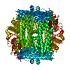
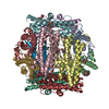
- Structure visualization
| Structure viewer | Molecule: MolmilJmol/JSmol |
|---|
- Downloads & links
-Download
| PDBx/mmCIF format | 2c2j.cif.gz | 49.7 KB | Display | PDBx/mmCIF format |
|---|---|---|---|---|
| PDB format | pdb2c2j.ent.gz | 35.2 KB | Display | PDB format |
| PDBx/mmJSON format | 2c2j.json.gz | Tree view | PDBx/mmJSON format | |
| Others |  Other downloads Other downloads |
-Validation report
| Arichive directory | https://data.pdbj.org/pub/pdb/validation_reports/c2/2c2jftp://data.pdbj.org/pub/pdb/validation_reports/c2/2c2j | HTTPS FTP |
|---|
-Related structure data

-Links
 PDBj
PDBj


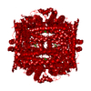
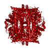
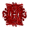
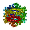
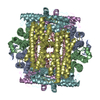
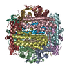
- Assembly
| Deposited unit | 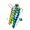
| |||||||||||||||
|---|---|---|---|---|---|---|---|---|---|---|---|---|---|---|---|---|
| 1 | x 12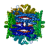
| |||||||||||||||
| Unit cell |
| |||||||||||||||
| Components on special symmetry positions |
|
-Components
| #1: Protein | Mass: 23289.715 Da / Num. of mol.: 1 Source method: isolated from a genetically manipulated source Details: DRB0092 WAS TRUNCATED - RESIDUE NUMBERING STARTS AFTER N-TERMINAL RESIDUE NUMBER 30 Source: (gene. exp.) DEINOCOCCUS RADIODURANS (radioresistant)Strain: R1 / Plasmid: PDEST14 / Production host: |
|---|---|
| #2: Chemical | ChemComp-FE /   Mass: 55.845 Da / Num. of mol.: 1 / Source method: obtained synthetically / Formula: Fe Mass: 55.845 Da / Num. of mol.: 1 / Source method: obtained synthetically / Formula: Fe |
| #3: Chemical | ChemComp-MG /   Mass: 24.305 Da / Num. of mol.: 1 / Source method: obtained synthetically / Formula: Mg Mass: 24.305 Da / Num. of mol.: 1 / Source method: obtained synthetically / Formula: Mg |
| #4: Water | ChemComp-HOH /  Mass: 18.015 Da / Num. of mol.: 128 / Source method: isolated from a natural source / Formula: H2O Mass: 18.015 Da / Num. of mol.: 128 / Source method: isolated from a natural source / Formula: H2O |
-Experimental details
-Experiment
| Experiment | Method: X-RAY DIFFRACTION / Number of used crystals: 1 |
|---|
- Sample preparation
| Crystal | Density Matthews: 2.5 Å3/Da / Density % sol: 54.3 % |
|---|---|
| Crystal grow | pH: 7.5 / Details: 0.01M MGCL2, 0.05M TRIS-HCL PH 7.5, 5% ISOPROPANOL |
-Data collection
| Diffraction | Mean temperature: 100 K |
|---|---|
| Diffraction source | Source: SYNCHROTRON / Site: ESRF  / Beamline: ID14-2 / Wavelength: 0.933 / Beamline: ID14-2 / Wavelength: 0.933 |
| Detector | Type: ADSC MARRESEARCH / Detector: CCD / Date: Mar 21, 2005 |
| Radiation | Protocol: SINGLE WAVELENGTH / Monochromatic (M) / Laue (L): M / Scattering type: x-ray |
| Radiation wavelength | Wavelength: 0.933 Å / Relative weight: 1 |
| Reflection | Resolution: 2.05→44.2 Å / Num. obs: 14773 / % possible obs: 100 % / Observed criterion σ(I): 1.5 / Redundancy: 21.1 % / Biso Wilson estimate: 29.8 Å2 / Rmerge(I) obs: 0.07 / Net I/σ(I): 40.4 |
| Reflection shell | Resolution: 2.05→2.1 Å / Redundancy: 18.6 % / Rmerge(I) obs: 0.5 / Mean I/σ(I) obs: 5.6 / % possible all: 100 |
- Processing
| Software |
| ||||||||||||||||||||||||||||||||||||||||||||||||||||||||||||||||||||||||||||||||||||||||||||||||||||||||||||||||||||||||||||||||||||||||||||||||||||||||||||||||||||||||||||||||||||||
|---|---|---|---|---|---|---|---|---|---|---|---|---|---|---|---|---|---|---|---|---|---|---|---|---|---|---|---|---|---|---|---|---|---|---|---|---|---|---|---|---|---|---|---|---|---|---|---|---|---|---|---|---|---|---|---|---|---|---|---|---|---|---|---|---|---|---|---|---|---|---|---|---|---|---|---|---|---|---|---|---|---|---|---|---|---|---|---|---|---|---|---|---|---|---|---|---|---|---|---|---|---|---|---|---|---|---|---|---|---|---|---|---|---|---|---|---|---|---|---|---|---|---|---|---|---|---|---|---|---|---|---|---|---|---|---|---|---|---|---|---|---|---|---|---|---|---|---|---|---|---|---|---|---|---|---|---|---|---|---|---|---|---|---|---|---|---|---|---|---|---|---|---|---|---|---|---|---|---|---|---|---|---|---|
| Refinement | Method to determine structure: SAD / Resolution: 2.05→88.74 Å / Cor.coef. Fo:Fc: 0.958 / SU B: 2.587 / SU ML: 0.073 / Cross valid method: THROUGHOUT / ESU R: 0.147 / Stereochemistry target values: MAXIMUM LIKELIHOOD Details: HYDROGENS HAVE BEEN ADDED IN THE RIDING POSITIONS. THE 41 N-TERMINAL AND 5 C-TERMINAL RESIDUES ARE MISSING IN THE STRUCTURE. WATERS 22, 31, 43 AND 126 HAVE OCCUPANCIES BELOW 1 BECAUSE OF ...Details: HYDROGENS HAVE BEEN ADDED IN THE RIDING POSITIONS. THE 41 N-TERMINAL AND 5 C-TERMINAL RESIDUES ARE MISSING IN THE STRUCTURE. WATERS 22, 31, 43 AND 126 HAVE OCCUPANCIES BELOW 1 BECAUSE OF THEIR LOCALISATION ON SYMMETRY AXIS. Z22 LIES ON A CELL EDGE AND A 2 FOLD AXIS Z43 LIES ON A CELL EDGE AND A 2 FOLD AXIS Z126 LIES ON A 3 FOLD AXIS
| ||||||||||||||||||||||||||||||||||||||||||||||||||||||||||||||||||||||||||||||||||||||||||||||||||||||||||||||||||||||||||||||||||||||||||||||||||||||||||||||||||||||||||||||||||||||
| Solvent computation | Ion probe radii: 0.8 Å / Shrinkage radii: 0.8 Å / VDW probe radii: 1.2 Å / Solvent model: MASK | ||||||||||||||||||||||||||||||||||||||||||||||||||||||||||||||||||||||||||||||||||||||||||||||||||||||||||||||||||||||||||||||||||||||||||||||||||||||||||||||||||||||||||||||||||||||
| Displacement parameters | Biso mean: 31.91 Å2 | ||||||||||||||||||||||||||||||||||||||||||||||||||||||||||||||||||||||||||||||||||||||||||||||||||||||||||||||||||||||||||||||||||||||||||||||||||||||||||||||||||||||||||||||||||||||
| Refinement step | Cycle: LAST / Resolution: 2.05→88.74 Å
| ||||||||||||||||||||||||||||||||||||||||||||||||||||||||||||||||||||||||||||||||||||||||||||||||||||||||||||||||||||||||||||||||||||||||||||||||||||||||||||||||||||||||||||||||||||||
| Refine LS restraints |
|