 Movie
Movie Controller

[English] 日本語
 Yorodumi
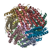
- PDB-1o9r: The X-ray crystal structure of Agrobacterium tumefaciens Dps, a m... -
+ Open data

- Basic information
| Entry | Database: PDB / ID: 1o9r | ||||||
|---|---|---|---|---|---|---|---|
| Title | The X-ray crystal structure of Agrobacterium tumefaciens Dps, a member of the family that protect DNA without binding | ||||||
 Components Components | AGROBACTERIUM TUMEFACIENS DPS | ||||||
 Keywords Keywords | IRON-BINDING PROTEIN / DNA PROTECTION FROM OXIDATIVE DAMAGE / DNA-BINDING / IRON- BINDING PROTEIN | ||||||
| Function / homology |  Function and homology information Function and homology informationOxidoreductases; Oxidizing metal ions / oxidoreductase activity, acting on metal ions / ferric iron binding / cytoplasm Similarity search - Function | ||||||
| Biological species |  AGROBACTERIUM TUMEFACIENS (bacteria) AGROBACTERIUM TUMEFACIENS (bacteria) | ||||||
| Method |  X-RAY DIFFRACTION / SYNCHROTRON / MOLECULAR REPLACEMENT / Resolution: 1.45 Å X-RAY DIFFRACTION / SYNCHROTRON / MOLECULAR REPLACEMENT / Resolution: 1.45 Å | ||||||
 Authors Authors | Ilari, A. / Ceci, P. / Chiancone, E. | ||||||
 Citation Citation | Journal: J.Biol.Chem. / Year: 2003 Title: The Dps Protein of Agrobacterium Tumefaciens Does not Bind to DNA But Protects It Toward Oxidative Cleavage: X-Ray Crystal Structure, Iron Binding, and Hydroxyl-Radical Scavenging Properties Authors: Ceci, P. / Ilari, A. / Falvo, E. / Chiancone, E. #1: Journal: J.Mol.Biol. / Year: 2002Title: Structure of the Neutrophil-Activating Protein from Helicobacter Pylori Authors: Zanotti, G. / Papinutto, E. / Dundon, W.G. / Battistutta, R. / Seveso, M. / Giudice, G.D. / Rappuoli, R. / Montecucco, C. #2: Journal: J.Biol.Chem. / Year: 2002Title: Structure of Two Iron-Binding Proteins from Bacillus Anthracis Authors: Papinutto, E. / Dundon, W.G. / Pitulis, N. / Battistutta, R. / Montecucco, C. / Zanotti, G. #3: Journal: Nat.Struct.Biol. / Year: 2000Title: The Dodecameric Ferritin from Listeria Innocua Contains a Novel Intersubunit Iron-Binding Site Authors: Ilari, A. / Stefanini, S. / Chiancone, E. / Tsernoglou, D. #4: Journal: Nat.Struct.Biol. / Year: 1998Title: The Crystal Structure of Dps, a Ferritin Homolog that Binds and Protects DNA Authors: Grant, R.A. / Filman, D.J. / Finkel, S.E. / Kolter, R. / Hogle, J.M. | ||||||
| History |
|
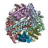
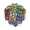
- Structure visualization
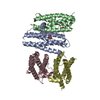
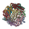
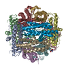
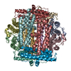
| Structure viewer | Molecule: MolmilJmol/JSmol |
|---|
- Downloads & links
-Download
| PDBx/mmCIF format | 1o9r.cif.gz | 205.2 KB | Display | PDBx/mmCIF format |
|---|---|---|---|---|
| PDB format | pdb1o9r.ent.gz | 166.8 KB | Display | PDB format |
| PDBx/mmJSON format | 1o9r.json.gz | Tree view | PDBx/mmJSON format | |
| Others |  Other downloads Other downloads |
-Validation report
| Arichive directory | https://data.pdbj.org/pub/pdb/validation_reports/o9/1o9rftp://data.pdbj.org/pub/pdb/validation_reports/o9/1o9r | HTTPS FTP |
|---|
-Related structure data
| Related structure data | 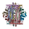 1dpsS S: Starting model for refinement |
|---|---|
| Similar structure data |

-Links
 PDBj
PDBj


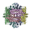
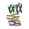
- Assembly
| Deposited unit | 
| ||||||||||||||||||||||||
|---|---|---|---|---|---|---|---|---|---|---|---|---|---|---|---|---|---|---|---|---|---|---|---|---|---|
| 1 | 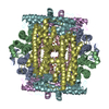
| ||||||||||||||||||||||||
| Unit cell |
| ||||||||||||||||||||||||
| Noncrystallographic symmetry (NCS) | NCS oper:
|
-Components
| #1: Protein | Mass: 17847.086 Da / Num. of mol.: 6 Source method: isolated from a genetically manipulated source Source: (gene. exp.) AGROBACTERIUM TUMEFACIENS (bacteria) / Strain: GV3101 / Plasmid: PET11-A / Production host: #2: Chemical | ChemComp-FE /   Mass: 55.845 Da / Num. of mol.: 6 / Source method: obtained synthetically / Formula: Fe Mass: 55.845 Da / Num. of mol.: 6 / Source method: obtained synthetically / Formula: Fe#3: Chemical |   Mass: 122.143 Da / Num. of mol.: 2 / Source method: obtained synthetically / Formula: C4H12NO3 / Comment: pH buffer*YM Mass: 122.143 Da / Num. of mol.: 2 / Source method: obtained synthetically / Formula: C4H12NO3 / Comment: pH buffer*YM#4: Chemical | ChemComp-EDO / |   Mass: 62.068 Da / Num. of mol.: 1 / Source method: obtained synthetically / Formula: C2H6O2 Mass: 62.068 Da / Num. of mol.: 1 / Source method: obtained synthetically / Formula: C2H6O2#5: Water | ChemComp-HOH / |  Mass: 18.015 Da / Num. of mol.: 741 / Source method: isolated from a natural source / Formula: H2O Mass: 18.015 Da / Num. of mol.: 741 / Source method: isolated from a natural source / Formula: H2O |
|---|
-Experimental details
-Experiment
| Experiment | Method: X-RAY DIFFRACTION |
|---|
- Sample preparation
| Crystal | Density Matthews: 2.26 Å3/Da / Density % sol: 45 % | ||||||||||||||||||||||||||||||||||||||||||
|---|---|---|---|---|---|---|---|---|---|---|---|---|---|---|---|---|---|---|---|---|---|---|---|---|---|---|---|---|---|---|---|---|---|---|---|---|---|---|---|---|---|---|---|
| Crystal grow | pH: 7.5 Details: HEPES 0.1 M, PH 7.0-7.8, ETHYLENE GLYCOL IN A RANGE BETWEEN 16%-24% V/V | ||||||||||||||||||||||||||||||||||||||||||
| Crystal grow | *PLUS Temperature: 293 K / Method: vapor diffusion, hanging drop | ||||||||||||||||||||||||||||||||||||||||||
| Components of the solutions | *PLUS
|
-Data collection
| Diffraction | Mean temperature: 100 K |
|---|---|
| Diffraction source | Source: SYNCHROTRON / Site: ELETTRA  / Beamline: 5.2R / Wavelength: 1.2 / Beamline: 5.2R / Wavelength: 1.2 |
| Radiation | Protocol: SINGLE WAVELENGTH / Monochromatic (M) / Laue (L): M / Scattering type: x-ray |
| Radiation wavelength | Wavelength: 1.2 Å / Relative weight: 1 |
| Reflection | Resolution: 1.45→50 Å / Num. obs: 179135 / % possible obs: 96 % / Observed criterion σ(I): 0.9 / Redundancy: 5 % / Biso Wilson estimate: 12.44 Å2 / Rmerge(I) obs: 0.053 |
| Reflection | *PLUS Lowest resolution: 50 Å / % possible obs: 96 % |
- Processing
| Software |
| ||||||||||||||||||||
|---|---|---|---|---|---|---|---|---|---|---|---|---|---|---|---|---|---|---|---|---|---|
| Refinement | Method to determine structure: MOLECULAR REPLACEMENT Starting model: PDB ENTRY 1DPS Resolution: 1.45→20 Å / Cross valid method: THROUGHOUT
| ||||||||||||||||||||
| Refinement step | Cycle: LAST / Resolution: 1.45→20 Å
| ||||||||||||||||||||
| Refinement | *PLUS Lowest resolution: 50 Å / Rfactor Rfree: 0.2 / Rfactor Rwork: 0.18 | ||||||||||||||||||||
| Solvent computation | *PLUS | ||||||||||||||||||||
| Displacement parameters | *PLUS | ||||||||||||||||||||
| Refine LS restraints | *PLUS
|
