 Movie
Movie Controller

[English] 日本語
 Yorodumi
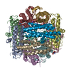
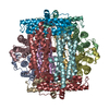
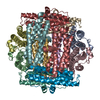
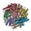
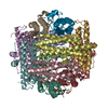
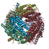
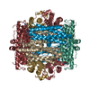
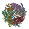
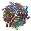
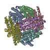
- PDB-1dps: THE CRYSTAL STRUCTURE OF DPS, A FERRITIN HOMOLOG THAT BINDS AND P... -
+ Open data

- Basic information
| Entry | Database: PDB / ID: 1dps | ||||||
|---|---|---|---|---|---|---|---|
| Title | THE CRYSTAL STRUCTURE OF DPS, A FERRITIN HOMOLOG THAT BINDS AND PROTECTS DNA | ||||||
 Components Components | DPS | ||||||
 Keywords Keywords | DNA BINDING PROTEIN / DNA-BINDING PROTEIN / FERRITIN / IRON SEQUESTRATION / STATIONARY PHASE / OXIDATIVE DAMAGE | ||||||
| Function / homology |  Function and homology information Function and homology informationDnaA-Dps complex / Oxidoreductases; Oxidizing metal ions / oxidoreductase activity, acting on metal ions / nucleoid / chromosome condensation / response to starvation / response to stress / ferric iron binding / negative regulation of DNA-templated DNA replication initiation / intracellular iron ion homeostasis ...DnaA-Dps complex / Oxidoreductases; Oxidizing metal ions / oxidoreductase activity, acting on metal ions / nucleoid / chromosome condensation / response to starvation / response to stress / ferric iron binding / negative regulation of DNA-templated DNA replication initiation / intracellular iron ion homeostasis / DNA binding / membrane / identical protein binding / cytoplasm Similarity search - Function | ||||||
| Biological species |  | ||||||
| Method |  X-RAY DIFFRACTION / SYNCHROTRON / MIR, NCS / Resolution: 1.6 Å X-RAY DIFFRACTION / SYNCHROTRON / MIR, NCS / Resolution: 1.6 Å | ||||||
 Authors Authors | Grant, R.A. / Filman, D.J. / Finkel, S.E. / Kolter, R. / Hogle, J.M. | ||||||
 Citation Citation | Journal: Nat.Struct.Biol. / Year: 1998 Title: The crystal structure of Dps, a ferritin homolog that binds and protects DNA. Authors: Grant, R.A. / Filman, D.J. / Finkel, S.E. / Kolter, R. / Hogle, J.M. | ||||||
| History |
|
- Structure visualization
| Structure viewer | Molecule: MolmilJmol/JSmol |
|---|
- Downloads & links
-Download
| PDBx/mmCIF format | 1dps.cif.gz | 383.9 KB | Display | PDBx/mmCIF format |
|---|---|---|---|---|
| PDB format | pdb1dps.ent.gz | 315.8 KB | Display | PDB format |
| PDBx/mmJSON format | 1dps.json.gz | Tree view | PDBx/mmJSON format | |
| Others |  Other downloads Other downloads |
-Validation report
| Arichive directory | https://data.pdbj.org/pub/pdb/validation_reports/dp/1dpsftp://data.pdbj.org/pub/pdb/validation_reports/dp/1dps | HTTPS FTP |
|---|
-Related structure data
| Similar structure data |
|---|

-Links
 PDBj
PDBj


- Assembly
| Deposited unit | 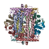
| ||||||||||||||||||||||||||||||||||||||||||||||||
|---|---|---|---|---|---|---|---|---|---|---|---|---|---|---|---|---|---|---|---|---|---|---|---|---|---|---|---|---|---|---|---|---|---|---|---|---|---|---|---|---|---|---|---|---|---|---|---|---|---|
| 1 |
| ||||||||||||||||||||||||||||||||||||||||||||||||
| Unit cell |
| ||||||||||||||||||||||||||||||||||||||||||||||||
| Noncrystallographic symmetry (NCS) | NCS oper:
|
-Components
| #1: Protein | Mass: 18736.359 Da / Num. of mol.: 12 / Mutation: S164C Source method: isolated from a genetically manipulated source Source: (gene. exp.) #2: Chemical | ChemComp-NA /   Mass: 22.990 Da / Num. of mol.: 12 / Source method: obtained synthetically / Formula: Na Mass: 22.990 Da / Num. of mol.: 12 / Source method: obtained synthetically / Formula: Na#3: Water | ChemComp-HOH / |  Mass: 18.015 Da / Num. of mol.: 1397 / Source method: isolated from a natural source / Formula: H2O Mass: 18.015 Da / Num. of mol.: 1397 / Source method: isolated from a natural source / Formula: H2O |
|---|
-Experimental details
-Experiment
| Experiment | Method: X-RAY DIFFRACTION / Number of used crystals: 4 |
|---|
- Sample preparation
| Crystal | Density Matthews: 2.4 Å3/Da / Density % sol: 53 % / Description: FUJI IMAGE PLATES FOR CHESS DATA |
|---|---|
| Crystal grow | pH: 8 Details: 1.55-1.7 M SODIUM FORMATE 13-16% PEG 8000 100 MM NACL 50 MM TRIS PH 8, pH 8.0 |
-Data collection
| Diffraction | Mean temperature: 113 K |
|---|---|
| Diffraction source | Source: SYNCHROTRON / Site: CHESS  / Beamline: F1 / Wavelength: 1.5418 / Beamline: F1 / Wavelength: 1.5418 |
| Detector | Type: MARRESEARCH / Detector: IMAGE PLATE / Details: MIRRORS |
| Radiation | Monochromatic (M) / Laue (L): M / Scattering type: x-ray |
| Radiation wavelength | Wavelength: 1.5418 Å / Relative weight: 1 |
| Reflection | Resolution: 1.6→20 Å / Num. obs: 1792266 / % possible obs: 92 % / Biso Wilson estimate: 20.2 Å2 / Rsym value: 0.046 / Net I/σ(I): 14.2 |
| Reflection shell | Resolution: 1.6→1.66 Å / Rmerge(I) obs: 0.052 / Mean I/σ(I) obs: 4.6 / Rsym value: 0.052 / % possible all: 80.1 |
- Processing
| Software |
| ||||||||||||||||||||||||||||||||||||||||||||||||||||||||||||
|---|---|---|---|---|---|---|---|---|---|---|---|---|---|---|---|---|---|---|---|---|---|---|---|---|---|---|---|---|---|---|---|---|---|---|---|---|---|---|---|---|---|---|---|---|---|---|---|---|---|---|---|---|---|---|---|---|---|---|---|---|---|
| Refinement | Method to determine structure: MIR, NCS / Resolution: 1.6→20 Å / Rfactor Rfree error: 0.001 / Data cutoff high absF: 10000000 / Data cutoff low absF: 0.001 / Cross valid method: THROUGHOUT / σ(F): 2 / Details: BULK SOLVENT MODEL USED
| ||||||||||||||||||||||||||||||||||||||||||||||||||||||||||||
| Displacement parameters | Biso mean: 18.3 Å2 | ||||||||||||||||||||||||||||||||||||||||||||||||||||||||||||
| Refine analyze |
| ||||||||||||||||||||||||||||||||||||||||||||||||||||||||||||
| Refinement step | Cycle: LAST / Resolution: 1.6→20 Å
| ||||||||||||||||||||||||||||||||||||||||||||||||||||||||||||
| Refine LS restraints |
| ||||||||||||||||||||||||||||||||||||||||||||||||||||||||||||
| LS refinement shell | Resolution: 1.6→1.7 Å / Rfactor Rfree error: 0.004 / Total num. of bins used: 6
| ||||||||||||||||||||||||||||||||||||||||||||||||||||||||||||
| Xplor file |
|
