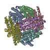
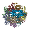
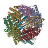
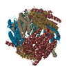
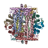
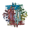
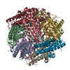
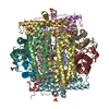
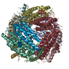
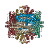
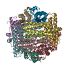
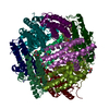
- PDB-1jts: DNA PROTECTION AND BINDING BY E. COLI DPS PROTEIN -
+
Open data
ID or keywords:
Loading...
-
Basic information
Entry
Database: PDB / ID: 1jts
Title
DNA PROTECTION AND BINDING BY E. COLI DPS PROTEIN
Components
DNA PROTECTION DURING STARVATION PROTEIN
Keywords
DNA BINDING PROTEIN / DODECAMER
Function / homology
Function and homology information
DnaA-Dps complex / Oxidoreductases; Oxidizing metal ions / oxidoreductase activity, acting on metal ions / nucleoid / chromosome condensation / response to starvation / response to stress / ferric iron binding / negative regulation of DNA-templated DNA replication initiation / intracellular iron ion homeostasis ...DnaA-Dps complex / Oxidoreductases; Oxidizing metal ions / oxidoreductase activity, acting on metal ions / nucleoid / chromosome condensation / response to starvation / response to stress / ferric iron binding / negative regulation of DNA-templated DNA replication initiation / intracellular iron ion homeostasis / DNA binding / membrane / identical protein binding / cytoplasm Similarity search - Function
DNA protection during starvation protein, gammaproteobacteria / Dps protein family signature 2. / Dps protein family signature 1. / DNA-binding protein Dps, conserved site / DNA-binding protein Dps / Ferritin, core subunit, four-helix bundle / Ferritin / Ferritin/DPS protein domain / Ferritin-like domain / Ferritin-like ...DNA protection during starvation protein, gammaproteobacteria / Dps protein family signature 2. / Dps protein family signature 1. / DNA-binding protein Dps, conserved site / DNA-binding protein Dps / Ferritin, core subunit, four-helix bundle / Ferritin / Ferritin/DPS protein domain / Ferritin-like domain / Ferritin-like / Ferritin-like superfamily / Up-down Bundle / Mainly Alpha Similarity search - Domain/homology
A: DNA PROTECTION DURING STARVATION PROTEIN B: DNA PROTECTION DURING STARVATION PROTEIN C: DNA PROTECTION DURING STARVATION PROTEIN D: DNA PROTECTION DURING STARVATION PROTEIN E: DNA PROTECTION DURING STARVATION PROTEIN F: DNA PROTECTION DURING STARVATION PROTEIN G: DNA PROTECTION DURING STARVATION PROTEIN H: DNA PROTECTION DURING STARVATION PROTEIN I: DNA PROTECTION DURING STARVATION PROTEIN J: DNA PROTECTION DURING STARVATION PROTEIN K: DNA PROTECTION DURING STARVATION PROTEIN L: DNA PROTECTION DURING STARVATION PROTEIN M: DNA PROTECTION DURING STARVATION PROTEIN N: DNA PROTECTION DURING STARVATION PROTEIN O: DNA PROTECTION DURING STARVATION PROTEIN P: DNA PROTECTION DURING STARVATION PROTEIN Q: DNA PROTECTION DURING STARVATION PROTEIN R: DNA PROTECTION DURING STARVATION PROTEIN S: DNA PROTECTION DURING STARVATION PROTEIN T: DNA PROTECTION DURING STARVATION PROTEIN U: DNA PROTECTION DURING STARVATION PROTEIN V: DNA PROTECTION DURING STARVATION PROTEIN W: DNA PROTECTION DURING STARVATION PROTEIN X: DNA PROTECTION DURING STARVATION PROTEIN hetero molecules
A: DNA PROTECTION DURING STARVATION PROTEIN B: DNA PROTECTION DURING STARVATION PROTEIN C: DNA PROTECTION DURING STARVATION PROTEIN D: DNA PROTECTION DURING STARVATION PROTEIN E: DNA PROTECTION DURING STARVATION PROTEIN F: DNA PROTECTION DURING STARVATION PROTEIN G: DNA PROTECTION DURING STARVATION PROTEIN H: DNA PROTECTION DURING STARVATION PROTEIN I: DNA PROTECTION DURING STARVATION PROTEIN J: DNA PROTECTION DURING STARVATION PROTEIN K: DNA PROTECTION DURING STARVATION PROTEIN L: DNA PROTECTION DURING STARVATION PROTEIN hetero molecules
M: DNA PROTECTION DURING STARVATION PROTEIN N: DNA PROTECTION DURING STARVATION PROTEIN O: DNA PROTECTION DURING STARVATION PROTEIN P: DNA PROTECTION DURING STARVATION PROTEIN Q: DNA PROTECTION DURING STARVATION PROTEIN R: DNA PROTECTION DURING STARVATION PROTEIN S: DNA PROTECTION DURING STARVATION PROTEIN T: DNA PROTECTION DURING STARVATION PROTEIN U: DNA PROTECTION DURING STARVATION PROTEIN V: DNA PROTECTION DURING STARVATION PROTEIN W: DNA PROTECTION DURING STARVATION PROTEIN X: DNA PROTECTION DURING STARVATION PROTEIN hetero molecules
In the structure databanks used in Yorodumi, some data are registered as the other names, "COVID-19 virus" and "2019-nCoV". Here are the details of the virus and the list of structure data.
Jan 31, 2019. EMDB accession codes are about to change! (news from PDBe EMDB page)
EMDB accession codes are about to change! (news from PDBe EMDB page)
The allocation of 4 digits for EMDB accession codes will soon come to an end. Whilst these codes will remain in use, new EMDB accession codes will include an additional digit and will expand incrementally as the available range of codes is exhausted. The current 4-digit format prefixed with “EMD-” (i.e. EMD-XXXX) will advance to a 5-digit format (i.e. EMD-XXXXX), and so on. It is currently estimated that the 4-digit codes will be depleted around Spring 2019, at which point the 5-digit format will come into force.
The EM Navigator/Yorodumi systems omit the EMD- prefix.
Related info.:Q: What is EMD? / ID/Accession-code notation in Yorodumi/EM Navigator
Yorodumi is a browser for structure data from EMDB, PDB, SASBDB, etc.
This page is also the successor to EM Navigator detail page, and also detail information page/front-end page for Omokage search.
The word "yorodu" (or yorozu) is an old Japanese word meaning "ten thousand". "mi" (miru) is to see.
Related info.:EMDB / PDB / SASBDB / Comparison of 3 databanks / Yorodumi Search / Aug 31, 2016. New EM Navigator & Yorodumi / Yorodumi Papers / Jmol/JSmol / Function and homology information / Changes in new EM Navigator and Yorodumi
 Movie
Movie Controller


 Open data
 Basic information
 Components
 Keywords
 Function and homology information
 X-RAY DIFFRACTION /
  Authors
 Citation
 Structure visualization
 Downloads & links
 Other downloads
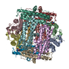


 PDBj
PDBj


 Assembly

 Mass: 122.143 Da / Num. of mol.: 24 / Source method: obtained synthetically / Formula: C4H12NO3 / Comment: pH buffer*YM
 Mass: 18.015 Da / Num. of mol.: 242 / Source method: isolated from a natural source / Formula: H2O
 Sample preparation
 / Beamline: X12B / Wavelength: 0.978
  Processing