 Movie
Movie Controller

[English] 日本語
 Yorodumi
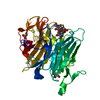
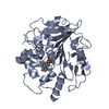
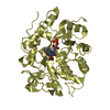
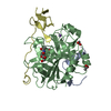
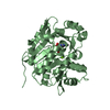
- PDB-6zeh: Structure of PP1-spectrin alpha II chimera [PP1(7-304) + linker (... -
+ Open data

- Basic information
| Entry | Database: PDB / ID: 6zeh | ||||||
|---|---|---|---|---|---|---|---|
| Title | Structure of PP1-spectrin alpha II chimera [PP1(7-304) + linker (G/S)x9 + spectrin alpha II (1025-1039)] bound to Phactr1 (516-580) | ||||||
 Components Components |
| ||||||
 Keywords Keywords | HYDROLASE / PP1 / Phosphatase / Phactr / RPEL | ||||||
| Function / homology |  Function and homology information Function and homology informationspectrin / regulation of glycogen catabolic process / positive regulation of termination of RNA polymerase II transcription, poly(A)-coupled / PTW/PP1 phosphatase complex / protein phosphatase type 1 complex / dendrite arborization / glycogen granule / RNA polymerase II promoter clearance / RNA polymerase II CTD heptapeptide repeat S5 phosphatase activity / protein phosphatase 1 binding ...spectrin / regulation of glycogen catabolic process / positive regulation of termination of RNA polymerase II transcription, poly(A)-coupled / PTW/PP1 phosphatase complex / protein phosphatase type 1 complex / dendrite arborization / glycogen granule / RNA polymerase II promoter clearance / RNA polymerase II CTD heptapeptide repeat S5 phosphatase activity / protein phosphatase 1 binding / cadherin binding involved in cell-cell adhesion / regulation of translational initiation in response to stress / actin filament capping / regulation of neuron migration / positive regulation of extrinsic apoptotic signaling pathway in absence of ligand / actomyosin structure organization / Nephrin family interactions / dephosphorylation / regulation of canonical Wnt signaling pathway / Sensory processing of sound by outer hair cells of the cochlea / Interaction between L1 and Ankyrins / Sensory processing of sound by inner hair cells of the cochlea / RHOV GTPase cycle / protein phosphatase inhibitor activity / cortical actin cytoskeleton / glycogen metabolic process / protein-serine/threonine phosphatase / stress fiber assembly / branching morphogenesis of an epithelial tube / Triglyceride catabolism / entrainment of circadian clock by photoperiod / Maturation of hRSV A proteins / protein serine/threonine phosphatase activity / phosphatase activity / telomere maintenance in response to DNA damage / RHOU GTPase cycle / phosphoprotein phosphatase activity / negative regulation of transcription elongation by RNA polymerase II / transition metal ion binding / DARPP-32 events / Caspase-mediated cleavage of cytoskeletal proteins / positive regulation of glycogen biosynthetic process / ribonucleoprotein complex binding / protein dephosphorylation / COPI-mediated anterograde transport / NCAM signaling for neurite out-growth / lung development / Downregulation of TGF-beta receptor signaling / cell projection / adherens junction / cell motility / circadian regulation of gene expression / positive regulation of transcription elongation by RNA polymerase II / regulation of circadian rhythm / cerebral cortex development / structural constituent of cytoskeleton / response to lead ion / specific granule lumen / actin filament binding / : / cell junction / tertiary granule lumen / extracellular vesicle / presynapse / actin binding / microtubule cytoskeleton / RAF/MAP kinase cascade / actin cytoskeleton organization / perikaryon / dendritic spine / calmodulin binding / protein stabilization / cadherin binding / iron ion binding / cell division / intracellular membrane-bounded organelle / calcium ion binding / synapse / Neutrophil degranulation / nucleolus / glutamatergic synapse / extracellular exosome / extracellular region / nucleoplasm / nucleus / membrane / plasma membrane / cytoplasm / cytosol Similarity search - Function | ||||||
| Biological species |  Homo sapiens (human) Homo sapiens (human) | ||||||
| Method |  X-RAY DIFFRACTION / SYNCHROTRON / MOLECULAR REPLACEMENT / Resolution: 1.3 Å X-RAY DIFFRACTION / SYNCHROTRON / MOLECULAR REPLACEMENT / Resolution: 1.3 Å | ||||||
 Authors Authors | Mouilleron, S. / Treisman, R. / Fedoryshchak, R. / Lee, R. / Butler, A.M. / Prechova, M. | ||||||
 Citation Citation | Journal: Elife / Year: 2020 Title: Molecular basis for substrate specificity of the Phactr1/PP1 phosphatase holoenzyme. Authors: Fedoryshchak, R.O. / Prechova, M. / Butler, A. / Lee, R. / O'Reilly, N. / Flynn, H.R. / Snijders, A.P. / Eder, N. / Ultanir, S. / Mouilleron, S. / Treisman, R. | ||||||
| History |
|
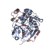
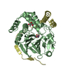
- Structure visualization
| Structure viewer | Molecule: MolmilJmol/JSmol |
|---|
- Downloads & links
-Download
| PDBx/mmCIF format | 6zeh.cif.gz | 566.1 KB | Display | PDBx/mmCIF format |
|---|---|---|---|---|
| PDB format | pdb6zeh.ent.gz | 389.7 KB | Display | PDB format |
| PDBx/mmJSON format | 6zeh.json.gz | Tree view | PDBx/mmJSON format | |
| Others |  Other downloads Other downloads |
-Validation report
| Summary document | 6zeh_validation.pdf.gz | 474 KB | Display | wwPDB validaton report |
|---|---|---|---|---|
| Full document | 6zeh_full_validation.pdf.gz | 477.8 KB | Display | |
| Data in XML | 6zeh_validation.xml.gz | 34.4 KB | Display | |
| Data in CIF | 6zeh_validation.cif.gz | 52 KB | Display | |
| Arichive directory | https://data.pdbj.org/pub/pdb/validation_reports/ze/6zehftp://data.pdbj.org/pub/pdb/validation_reports/ze/6zeh | HTTPS FTP |
-Related structure data
| Related structure data | 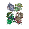 6zeeC  6zefC  6zegC  6zeiC  6zejC  4movS S: Starting model for refinement C: citing same article ( |
|---|---|
| Similar structure data |


-Links
 PDBj
PDBj


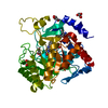
- Assembly
| Deposited unit | 
| ||||||||||
|---|---|---|---|---|---|---|---|---|---|---|---|
| 1 | 
| ||||||||||
| 2 | 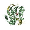
| ||||||||||
| Unit cell |
|
-Components
-Protein , 2 types, 4 molecules ABCD
| #1: Protein | Mass: 37558.555 Da / Num. of mol.: 2 / Mutation: N-terminal Vector derived sequence GHMGS Source method: isolated from a genetically manipulated source Source: (gene. exp.) Homo sapiens (human) / Gene: PPP1CA, PPP1A, SPTAN1, NEAS, SPTA2 / Production host:  References: UniProt: P62136, UniProt: Q13813, protein-serine/threonine phosphatase #2: Protein | Mass: 8257.345 Da / Num. of mol.: 2 / Mutation: N-terminal Vector derived sequence GPLGS Source method: isolated from a genetically manipulated source Source: (gene. exp.) Homo sapiens (human) / Gene: PHACTR1, hCG_1818446 / Production host: |
|---|
-Non-polymers , 4 types, 662 molecules 


| #3: Chemical | ChemComp-MN /  Mass: 54.938 Da / Num. of mol.: 4 / Source method: obtained synthetically / Formula: Mn Mass: 54.938 Da / Num. of mol.: 4 / Source method: obtained synthetically / Formula: Mn#4: Chemical |  Mass: 94.971 Da / Num. of mol.: 2 / Source method: isolated from a natural source / Formula: PO4 Mass: 94.971 Da / Num. of mol.: 2 / Source method: isolated from a natural source / Formula: PO4#5: Chemical | ChemComp-SO4 / |  Mass: 96.063 Da / Num. of mol.: 1 / Source method: obtained synthetically / Formula: SO4 Mass: 96.063 Da / Num. of mol.: 1 / Source method: obtained synthetically / Formula: SO4#6: Water | ChemComp-HOH / | Mass: 18.015 Da / Num. of mol.: 655 / Source method: isolated from a natural source / Formula: H2O |
|---|
-Details
| Has ligand of interest | N |
|---|
-Experimental details
-Experiment
| Experiment | Method: X-RAY DIFFRACTION / Number of used crystals: 1 |
|---|
- Sample preparation
| Crystal | Density Matthews: 2.26 Å3/Da / Density % sol: 45.69 % |
|---|---|
| Crystal grow | Temperature: 293 K / Method: vapor diffusion, sitting drop / pH: 8.5 Details: 20% (w/v) polyethylene glycol 3350, 0.2 M NaI and 0.1 M BIS-Tris propane pH 8.5 |
-Data collection
| Diffraction | Mean temperature: 100 K / Serial crystal experiment: N |
|---|---|
| Diffraction source | Source: SYNCHROTRON / Site: Diamond  / Beamline: I24 / Wavelength: 0.968 Å / Beamline: I24 / Wavelength: 0.968 Å |
| Detector | Type: DECTRIS PILATUS3 S 6M / Detector: PIXEL / Date: Jun 23, 2019 |
| Radiation | Protocol: SINGLE WAVELENGTH / Monochromatic (M) / Laue (L): M / Scattering type: x-ray |
| Radiation wavelength | Wavelength: 0.968 Å / Relative weight: 1 |
| Reflection | Resolution: 1.3→69.29 Å / Num. obs: 196809 / % possible obs: 99.08 % / Redundancy: 6.5 % / Biso Wilson estimate: 11.2 Å2 / CC1/2: 0.994 / Rmerge(I) obs: 0.1379 / Rpim(I) all: 0.058 / Rrim(I) all: 0.15 / Net I/σ(I): 5.89 |
| Reflection shell | Resolution: 1.3→1.34 Å / Rmerge(I) obs: 1.78 / Mean I/σ(I) obs: 1.12 / Num. unique obs: 123745 / CC1/2: 0.475 / Rpim(I) all: 0.75 / Rrim(I) all: 1.94 |
- Processing
| Software |
| |||||||||||||||||||||||||||||||||||||||||||||||||||||||||||||||||||||||||||||||||||||||||||||||||||||||||||||||||||||||||||||||||||||||||||||||||||||||||||||||||||||||||||||||||||||||||||||||||||||||||||||||||||||||||
|---|---|---|---|---|---|---|---|---|---|---|---|---|---|---|---|---|---|---|---|---|---|---|---|---|---|---|---|---|---|---|---|---|---|---|---|---|---|---|---|---|---|---|---|---|---|---|---|---|---|---|---|---|---|---|---|---|---|---|---|---|---|---|---|---|---|---|---|---|---|---|---|---|---|---|---|---|---|---|---|---|---|---|---|---|---|---|---|---|---|---|---|---|---|---|---|---|---|---|---|---|---|---|---|---|---|---|---|---|---|---|---|---|---|---|---|---|---|---|---|---|---|---|---|---|---|---|---|---|---|---|---|---|---|---|---|---|---|---|---|---|---|---|---|---|---|---|---|---|---|---|---|---|---|---|---|---|---|---|---|---|---|---|---|---|---|---|---|---|---|---|---|---|---|---|---|---|---|---|---|---|---|---|---|---|---|---|---|---|---|---|---|---|---|---|---|---|---|---|---|---|---|---|---|---|---|---|---|---|---|---|---|---|---|---|---|---|---|---|
| Refinement | Method to determine structure: MOLECULAR REPLACEMENT Starting model: 4MOV Resolution: 1.3→69.29 Å / SU ML: 0.123 / Cross valid method: FREE R-VALUE / σ(F): 1.35 / Phase error: 20.9272 Stereochemistry target values: GeoStd + Monomer Library + CDL v1.2
| |||||||||||||||||||||||||||||||||||||||||||||||||||||||||||||||||||||||||||||||||||||||||||||||||||||||||||||||||||||||||||||||||||||||||||||||||||||||||||||||||||||||||||||||||||||||||||||||||||||||||||||||||||||||||
| Solvent computation | Shrinkage radii: 0.9 Å / VDW probe radii: 1.11 Å / Solvent model: FLAT BULK SOLVENT MODEL | |||||||||||||||||||||||||||||||||||||||||||||||||||||||||||||||||||||||||||||||||||||||||||||||||||||||||||||||||||||||||||||||||||||||||||||||||||||||||||||||||||||||||||||||||||||||||||||||||||||||||||||||||||||||||
| Displacement parameters | Biso mean: 19.08 Å2 | |||||||||||||||||||||||||||||||||||||||||||||||||||||||||||||||||||||||||||||||||||||||||||||||||||||||||||||||||||||||||||||||||||||||||||||||||||||||||||||||||||||||||||||||||||||||||||||||||||||||||||||||||||||||||
| Refinement step | Cycle: LAST / Resolution: 1.3→69.29 Å
| |||||||||||||||||||||||||||||||||||||||||||||||||||||||||||||||||||||||||||||||||||||||||||||||||||||||||||||||||||||||||||||||||||||||||||||||||||||||||||||||||||||||||||||||||||||||||||||||||||||||||||||||||||||||||
| Refine LS restraints |
| |||||||||||||||||||||||||||||||||||||||||||||||||||||||||||||||||||||||||||||||||||||||||||||||||||||||||||||||||||||||||||||||||||||||||||||||||||||||||||||||||||||||||||||||||||||||||||||||||||||||||||||||||||||||||
| LS refinement shell |
|
