Type: MARMOSAIC 225 mm CCD / Detector: CCD / Date: Jun 2, 2011
Radiation
Protocol: SINGLE WAVELENGTH / Monochromatic (M) / Laue (L): M / Scattering type: x-ray
Radiation wavelength
Wavelength: 0.91502 Å / Relative weight: 1
Reflection
Resolution: 2.5→156.59 Å / Num. all: 22043 / Num. obs: 22043 / % possible obs: 99.9 % / Redundancy: 4.7 % / Rsym value: 0.08 / Net I/σ(I): 10.9
Reflection shell
Diffraction-ID: 1
Resolution (Å)
Redundancy (%)
Rmerge(I) obs
Mean I/σ(I) obs
Num. measured all
Num. unique all
Rpim(I) all
Rsym value
Net I/σ(I) obs
% possible all
2.5-2.64
4.5
0.262
2.9
14131
3143
0.154
0.262
4.5
100
2.64-2.8
4.7
0.206
3.6
14149
3021
0.12
0.206
5.6
100
2.8-2.99
4.8
0.141
5.3
13409
2803
0.081
0.141
7.6
100
2.99-3.23
4.8
0.097
7.7
12650
2640
0.056
0.097
9.9
100
3.23-3.54
4.8
0.069
10.3
11702
2435
0.039
0.069
13.2
100
3.54-3.95
4.8
0.056
12.6
10541
2218
0.031
0.056
15.4
100
3.95-4.56
4.7
0.055
11.7
9286
1965
0.03
0.055
16.9
100
4.56-5.59
4.6
0.067
8.6
7866
1699
0.036
0.067
16.5
100
5.59-7.91
4.5
0.054
10.6
6015
1329
0.031
0.054
15
99.8
7.91-52.197
4.2
0.047
10.2
3287
790
0.027
0.047
18.4
98.8
-
Phasing
Phasing
Method: molecular replacement
Phasing MR
Highest resolution
Lowest resolution
Rotation
3.5 Å
51.04 Å
Translation
3.5 Å
51.04 Å
-
Processing
Software
Name
Version
Classification
NB
MOSFLM
datareduction
SCALA
3.3.16
datascaling
MOLREP
phasing
REFMAC
refinement
PDB_EXTRACT
3.14
dataextraction
Refinement
Method to determine structure: MOLECULAR REPLACEMENT / Resolution: 2.5→156.59 Å / Cor.coef. Fo:Fc: 0.93 / Cor.coef. Fo:Fc free: 0.869 / SU B: 11.615 / SU ML: 0.272 / Cross valid method: THROUGHOUT / ESU R Free: 0.339 / Stereochemistry target values: MAXIMUM LIKELIHOOD / Details: HYDROGENS HAVE BEEN ADDED IN THE RIDING POSITIONS
Rfactor
Num. reflection
% reflection
Selection details
Rfree
0.2628
1128
5.1 %
RANDOM
Rwork
0.19611
-
-
-
obs
0.19962
20803
99.57 %
-
Solvent computation
Ion probe radii: 0.8 Å / Shrinkage radii: 0.8 Å / VDW probe radii: 1.2 Å / Solvent model: BABINET MODEL WITH MASK
 Movie
Movie Controller


 Open data
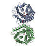
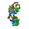
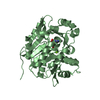
 Basic information
 Components
 Keywords
 Function and homology information
 Homo sapiens (human)
 X-RAY DIFFRACTION /
  Authors
 Citation
 Structure visualization
 Downloads & links
 Other downloads
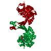


 PDBj
PDBj
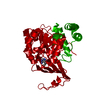
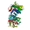
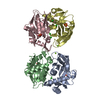
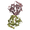
 Assembly


 Mass: 24.305 Da / Num. of mol.: 4 / Source method: obtained synthetically / Formula: Mg
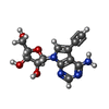
 Mass: 290.275 Da / Num. of mol.: 3 / Source method: obtained synthetically / Formula: C13H14N4O4
 Mass: 18.015 Da / Num. of mol.: 41 / Source method: isolated from a natural source / Formula: H2O
 Sample preparation
 / Beamline: 14.1 / Wavelength: 0.91502 Å
 Processing