 Movie
Movie Controller

[English] 日本語
 Yorodumi
- PDB-6uy4: Crystal structure of dihydroorotate dehydrogenase from Schistosom... -
+ Open data

- Basic information
| Entry | Database: PDB / ID: 6uy4 | |||||||||
|---|---|---|---|---|---|---|---|---|---|---|
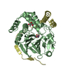
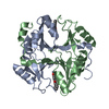
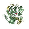
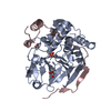
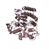
| Title | Crystal structure of dihydroorotate dehydrogenase from Schistosoma mansoni | |||||||||
 Components Components | Dihydroorotate dehydrogenase | |||||||||
 Keywords Keywords | OXIDOREDUCTASE/INHIBITOR / SmDHODH / class 2 dihydroorotate dehydrogenase / Schistosoma mansoni / FLAVOPROTEIN / OXIDOREDUCTASE-INHIBITOR complex | |||||||||
| Function / homology |  Function and homology information Function and homology informationdihydroorotate dehydrogenase (quinone) / dihydroorotate dehydrogenase (quinone) activity / 'de novo' UMP biosynthetic process / 'de novo' pyrimidine nucleobase biosynthetic process / mitochondrial inner membrane / nucleotide binding Similarity search - Function | |||||||||
| Biological species |  | |||||||||
| Method |  X-RAY DIFFRACTION / SYNCHROTRON / MOLECULAR REPLACEMENT / Resolution: 2.796 Å X-RAY DIFFRACTION / SYNCHROTRON / MOLECULAR REPLACEMENT / Resolution: 2.796 Å | |||||||||
 Authors Authors | Mori, R.M. / Zapata, L.C.C. / Nonato, M.C. | |||||||||
| Funding support |  Brazil, 2items Brazil, 2items
| |||||||||
 Citation Citation | Journal: Febs J. / Year: 2021 Title: Structural basis for the function and inhibition of dihydroorotate dehydrogenase from Schistosoma mansoni. Authors: de Mori, R.M. / Aleixo, M.A.A. / Zapata, L.C.C. / Calil, F.A. / Emery, F.S. / Nonato, M.C. | |||||||||
| History |
|
- Structure visualization
| Structure viewer | Molecule: MolmilJmol/JSmol |
|---|
- Downloads & links
-Download
| PDBx/mmCIF format | 6uy4.cif.gz | 85.7 KB | Display | PDBx/mmCIF format |
|---|---|---|---|---|
| PDB format | pdb6uy4.ent.gz | 62.3 KB | Display | PDB format |
| PDBx/mmJSON format | 6uy4.json.gz | Tree view | PDBx/mmJSON format | |
| Others |  Other downloads Other downloads |
-Validation report
| Arichive directory | https://data.pdbj.org/pub/pdb/validation_reports/uy/6uy4ftp://data.pdbj.org/pub/pdb/validation_reports/uy/6uy4 | HTTPS FTP |
|---|
-Related structure data
| Related structure data |  6uoy S: Starting model for refinement |
|---|---|
| Similar structure data |


-Links
 PDBj
PDBj- Assembly
| Deposited unit | 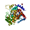
| ||||||||
|---|---|---|---|---|---|---|---|---|---|
| 1 |
| ||||||||
| Unit cell |
|
-Components
| #1: Protein | Mass: 41342.180 Da / Num. of mol.: 1 Source method: isolated from a genetically manipulated source Source: (gene. exp.)  References: UniProt: G4VFD7, dihydroorotate dehydrogenase (fumarate) | ||||||
|---|---|---|---|---|---|---|---|
| #2: Chemical | ChemComp-QLA /   Mass: 283.254 Da / Num. of mol.: 1 / Source method: obtained synthetically / Formula: C16H10FNO3 / Feature type: SUBJECT OF INVESTIGATION Mass: 283.254 Da / Num. of mol.: 1 / Source method: obtained synthetically / Formula: C16H10FNO3 / Feature type: SUBJECT OF INVESTIGATION | ||||||
| #3: Chemical | ChemComp-GOL /   Mass: 92.094 Da / Num. of mol.: 4 / Source method: obtained synthetically / Formula: C3H8O3 Mass: 92.094 Da / Num. of mol.: 4 / Source method: obtained synthetically / Formula: C3H8O3#4: Chemical | ChemComp-FMN / |   Mass: 456.344 Da / Num. of mol.: 1 / Source method: obtained synthetically / Formula: C17H21N4O9P Mass: 456.344 Da / Num. of mol.: 1 / Source method: obtained synthetically / Formula: C17H21N4O9P#5: Water | ChemComp-HOH / |  Mass: 18.015 Da / Num. of mol.: 45 / Source method: isolated from a natural source / Formula: H2O Mass: 18.015 Da / Num. of mol.: 45 / Source method: isolated from a natural source / Formula: H2OHas ligand of interest | Y | |
-Experimental details
-Experiment
| Experiment | Method: X-RAY DIFFRACTION / Number of used crystals: 1 |
|---|
- Sample preparation
| Crystal | Density Matthews: 3.45 Å3/Da / Density % sol: 64.4 % / Description: Yellow cube |
|---|---|
| Crystal grow | Temperature: 294 K / Method: vapor diffusion, sitting drop / Details: 0.1 MES pH 6.5; 1.4 M CITRIC ACID |
-Data collection
| Diffraction | Mean temperature: 100 K / Serial crystal experiment: N |
|---|---|
| Diffraction source | Source: SYNCHROTRON / Site: SOLEIL  / Beamline: PROXIMA 2 / Wavelength: 0.98 Å / Beamline: PROXIMA 2 / Wavelength: 0.98 Å |
| Detector | Type: DECTRIS EIGER X 9M / Detector: PIXEL / Date: Jun 26, 2019 |
| Radiation | Monochromator: Cryogenically colled channel cut crystal / Protocol: SINGLE WAVELENGTH / Monochromatic (M) / Laue (L): M / Scattering type: x-ray |
| Radiation wavelength | Wavelength: 0.98 Å / Relative weight: 1 |
| Reflection | Resolution: 2.796→47 Å / Num. obs: 14354 / % possible obs: 99.83 % / Redundancy: 39.8 % / CC1/2: 0.997 / Net I/σ(I): 15.2 |
| Reflection shell | Resolution: 2.8→2.95 Å / Num. unique obs: 2047 / CC1/2: 0.404 |
- Processing
| Software |
| ||||||||||||||||||||||||||||||||||||
|---|---|---|---|---|---|---|---|---|---|---|---|---|---|---|---|---|---|---|---|---|---|---|---|---|---|---|---|---|---|---|---|---|---|---|---|---|---|
| Refinement | Method to determine structure: MOLECULAR REPLACEMENT Starting model: 6UOY 6uoy Resolution: 2.796→46.995 Å / SU ML: 0.45 / Cross valid method: THROUGHOUT / σ(F): 1.33 / Phase error: 29.05 / Stereochemistry target values: ML
| ||||||||||||||||||||||||||||||||||||
| Solvent computation | Shrinkage radii: 0.9 Å / VDW probe radii: 1.11 Å / Solvent model: FLAT BULK SOLVENT MODEL | ||||||||||||||||||||||||||||||||||||
| Displacement parameters | Biso max: 172.68 Å2 / Biso mean: 79.8748 Å2 / Biso min: 51.19 Å2 | ||||||||||||||||||||||||||||||||||||
| Refinement step | Cycle: final / Resolution: 2.796→46.995 Å
| ||||||||||||||||||||||||||||||||||||
| LS refinement shell | Refine-ID: X-RAY DIFFRACTION / Rfactor Rfree error: 0
|
