 Movie
Movie Controller

[English] 日本語
 Yorodumi
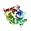
- PDB-6n1b: Crystal structure of an N-acetylgalactosamine deacetylase from F.... -
+ Open data

- Basic information
| Entry | Database: PDB / ID: 6n1b | |||||||||
|---|---|---|---|---|---|---|---|---|---|---|
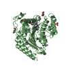
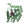
| Title | Crystal structure of an N-acetylgalactosamine deacetylase from F. plautii in complex with blood group B trisaccharide | |||||||||
 Components Components | Carbohydrate-binding protein | |||||||||
 Keywords Keywords | HYDROLASE / N-acetylgalactosamine deacetylase / A Blood antigen / CAZy | |||||||||
| Function / homology |  Function and homology information Function and homology informationmetabolic process / hydrolase activity, acting on glycosyl bonds / metal ion binding Similarity search - Function | |||||||||
| Biological species |  Flavonifractor plautii (bacteria) Flavonifractor plautii (bacteria) | |||||||||
| Method |  X-RAY DIFFRACTION / SYNCHROTRON / MOLECULAR REPLACEMENT / Resolution: 1.3 Å X-RAY DIFFRACTION / SYNCHROTRON / MOLECULAR REPLACEMENT / Resolution: 1.3 Å | |||||||||
 Authors Authors | Sim, L. / Rahfeld, P. / Withers, S.G. | |||||||||
| Funding support |  Canada, 1items Canada, 1items
| |||||||||
 Citation Citation | Journal: Nat Microbiol / Year: 2019 Title: An enzymatic pathway in the human gut microbiome that converts A to universal O type blood. Authors: Rahfeld, P. / Sim, L. / Moon, H. / Constantinescu, I. / Morgan-Lang, C. / Hallam, S.J. / Kizhakkedathu, J.N. / Withers, S.G. | |||||||||
| History |
|
- Structure visualization
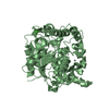
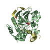
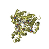
| Structure viewer | Molecule: MolmilJmol/JSmol |
|---|
- Downloads & links
-Download
| PDBx/mmCIF format | 6n1b.cif.gz | 108.1 KB | Display | PDBx/mmCIF format |
|---|---|---|---|---|
| PDB format | pdb6n1b.ent.gz | 76 KB | Display | PDB format |
| PDBx/mmJSON format | 6n1b.json.gz | Tree view | PDBx/mmJSON format | |
| Others |  Other downloads Other downloads |
-Validation report
| Arichive directory | https://data.pdbj.org/pub/pdb/validation_reports/n1/6n1bftp://data.pdbj.org/pub/pdb/validation_reports/n1/6n1b | HTTPS FTP |
|---|
-Related structure data
| Related structure data |  6n1aSC S: Starting model for refinement C: citing same article ( |
|---|---|
| Similar structure data |


-Links
 PDBj
PDBj


- Assembly
| Deposited unit | 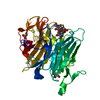
| ||||||||
|---|---|---|---|---|---|---|---|---|---|
| 1 |
| ||||||||
| Unit cell |
|
-Components
| #1: Protein | Mass: 55804.824 Da / Num. of mol.: 1 Source method: isolated from a genetically manipulated source Source: (gene. exp.) Flavonifractor plautii (bacteria) / Gene: A4U99_12080 / Production host: | ||||
|---|---|---|---|---|---|
| #2: Polysaccharide | alpha-L-fucopyranose-(1-2)-[alpha-D-galactopyranose-(1-3)]beta-D-galactopyranose Source method: isolated from a genetically manipulated source | ||||
| #3: Chemical | ChemComp-CA /   Mass: 40.078 Da / Num. of mol.: 4 / Source method: obtained synthetically / Formula: Ca Mass: 40.078 Da / Num. of mol.: 4 / Source method: obtained synthetically / Formula: Ca#4: Chemical |   Mass: 92.094 Da / Num. of mol.: 3 Mass: 92.094 Da / Num. of mol.: 3Source method: isolated from a genetically manipulated source Formula: C3H8O3 #5: Water | ChemComp-HOH / |  Mass: 18.015 Da / Num. of mol.: 359 / Source method: isolated from a natural source / Formula: H2O Mass: 18.015 Da / Num. of mol.: 359 / Source method: isolated from a natural source / Formula: H2O |
-Experimental details
-Experiment
| Experiment | Method: X-RAY DIFFRACTION / Number of used crystals: 1 |
|---|
- Sample preparation
| Crystal | Density Matthews: 1.68 Å3/Da / Density % sol: 26.84 % |
|---|---|
| Crystal grow | Temperature: 293 K / Method: vapor diffusion, hanging drop / Details: 0.2 M CaCl2, 0.1 M MES pH 6.0, 18% PEG 4000 |
-Data collection
| Diffraction | Mean temperature: 100 K / Serial crystal experiment: N |
|---|---|
| Diffraction source | Source: SYNCHROTRON / Site: CLSI / Beamline: 08ID-1 / Wavelength: 0.979 Å |
| Detector | Type: DECTRIS PILATUS3 S 6M / Detector: PIXEL / Date: Mar 12, 2018 |
| Radiation | Protocol: SINGLE WAVELENGTH / Monochromatic (M) / Laue (L): M / Scattering type: x-ray |
| Radiation wavelength | Wavelength: 0.979 Å / Relative weight: 1 |
| Reflection | Resolution: 1.3→46.4 Å / Num. obs: 90596 / % possible obs: 97.3 % / Redundancy: 5.5 % / CC1/2: 1 / Rpim(I) all: 0.024 / Net I/av σ(I): 16.3 / Net I/σ(I): 16.3 |
| Reflection shell | Resolution: 1.3→1.32 Å / Redundancy: 1.9 % / Mean I/σ(I) obs: 1.9 / Num. unique obs: 3142 / CC1/2: 0.76 / Rpim(I) all: 0.259 / % possible all: 69.1 |
- Processing
| Software |
| |||||||||||||||||||||||||||||||||||||||||||||||||||||||||||||||||||||||||||
|---|---|---|---|---|---|---|---|---|---|---|---|---|---|---|---|---|---|---|---|---|---|---|---|---|---|---|---|---|---|---|---|---|---|---|---|---|---|---|---|---|---|---|---|---|---|---|---|---|---|---|---|---|---|---|---|---|---|---|---|---|---|---|---|---|---|---|---|---|---|---|---|---|---|---|---|---|
| Refinement | Method to determine structure: MOLECULAR REPLACEMENT Starting model: 6N1A Resolution: 1.3→46.4 Å / Cor.coef. Fo:Fc: 0.975 / Cor.coef. Fo:Fc free: 0.97 / Cross valid method: THROUGHOUT / σ(F): 0 / ESU R: 0.043 / ESU R Free: 0.044 Details: HYDROGENS HAVE BEEN ADDED IN THE RIDING POSITIONS U VALUES : REFINED INDIVIDUALLY
| |||||||||||||||||||||||||||||||||||||||||||||||||||||||||||||||||||||||||||
| Solvent computation | Ion probe radii: 0.8 Å / Shrinkage radii: 0.8 Å / VDW probe radii: 1.2 Å | |||||||||||||||||||||||||||||||||||||||||||||||||||||||||||||||||||||||||||
| Displacement parameters | Biso max: 111.72 Å2 / Biso mean: 13.372 Å2 / Biso min: 6.35 Å2
| |||||||||||||||||||||||||||||||||||||||||||||||||||||||||||||||||||||||||||
| Refinement step | Cycle: final / Resolution: 1.3→46.4 Å
| |||||||||||||||||||||||||||||||||||||||||||||||||||||||||||||||||||||||||||
| Refine LS restraints |
| |||||||||||||||||||||||||||||||||||||||||||||||||||||||||||||||||||||||||||
| LS refinement shell | Resolution: 1.3→1.334 Å / Total num. of bins used: 20
|
