 Movie
Movie Controller


+ Open data

- Basic information
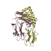
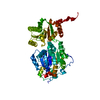
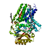
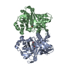
| Entry | Database: PDB / ID: 6z1o | ||||||
|---|---|---|---|---|---|---|---|
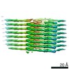
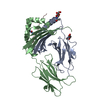
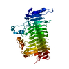
| Title | AL amyloid fibril from a lambda 3 light chain in conformation A | ||||||
 Components Components | lambda 3 immunoglobulin light chain fragment, residues 2-116 | ||||||
 Keywords Keywords | IMMUNE SYSTEM / amyloid / antibody / systemic amyloidosis / light chain | ||||||
| Function / homology |  Function and homology information Function and homology informationCD22 mediated BCR regulation / Fc epsilon receptor (FCERI) signaling / Classical antibody-mediated complement activation / Initial triggering of complement / FCGR activation / Role of LAT2/NTAL/LAB on calcium mobilization / Role of phospholipids in phagocytosis / immunoglobulin complex / Scavenging of heme from plasma / antigen binding ...CD22 mediated BCR regulation / Fc epsilon receptor (FCERI) signaling / Classical antibody-mediated complement activation / Initial triggering of complement / FCGR activation / Role of LAT2/NTAL/LAB on calcium mobilization / Role of phospholipids in phagocytosis / immunoglobulin complex / Scavenging of heme from plasma / antigen binding / FCERI mediated Ca+2 mobilization / FCGR3A-mediated IL10 synthesis / Regulation of Complement cascade / Antigen activates B Cell Receptor (BCR) leading to generation of second messengers / Cell surface interactions at the vascular wall / FCGR3A-mediated phagocytosis / FCERI mediated MAPK activation / Regulation of actin dynamics for phagocytic cup formation / Immunoregulatory interactions between a Lymphoid and a non-Lymphoid cell / FCERI mediated NF-kB activation / Potential therapeutics for SARS / adaptive immune response / immune response / extracellular exosome / extracellular region / plasma membrane Similarity search - Function | ||||||
| Biological species |  Homo sapiens (human) Homo sapiens (human) | ||||||
| Method | ELECTRON MICROSCOPY / helical reconstruction / cryo EM / Resolution: 3.2 Å | ||||||
 Authors Authors | Radamaker, L. / Fandrich, M. | ||||||
| Funding support |  Germany, 1items Germany, 1items
| ||||||
 Citation Citation | Journal: Nat Commun / Year: 2021 Title: Cryo-EM reveals structural breaks in a patient-derived amyloid fibril from systemic AL amyloidosis. Authors: Lynn Radamaker / Julian Baur / Stefanie Huhn / Christian Haupt / Ute Hegenbart / Stefan Schönland / Akanksha Bansal / Matthias Schmidt / Marcus Fändrich / Abstract: Systemic AL amyloidosis is a debilitating and potentially fatal disease that arises from the misfolding and fibrillation of immunoglobulin light chains (LCs). The disease is patient-specific with ...Systemic AL amyloidosis is a debilitating and potentially fatal disease that arises from the misfolding and fibrillation of immunoglobulin light chains (LCs). The disease is patient-specific with essentially each patient possessing a unique LC sequence. In this study, we present two ex vivo fibril structures of a λ3 LC. The fibrils were extracted from the explanted heart of a patient (FOR005) and consist of 115-residue fibril proteins, mainly from the LC variable domain. The fibril structures imply that a 180° rotation around the disulfide bond and a major unfolding step are necessary for fibrils to form. The two fibril structures show highly similar fibril protein folds, differing in only a 12-residue segment. Remarkably, the two structures do not represent separate fibril morphologies, as they can co-exist at different z-axial positions within the same fibril. Our data imply the presence of structural breaks at the interface of the two structural forms. | ||||||
| History |
|
- Structure visualization
| Movie |
Movie viewer |
|---|---|
| Structure viewer | Molecule: MolmilJmol/JSmol |
- Downloads & links
-Download
| PDBx/mmCIF format | 6z1o.cif.gz | 165.8 KB | Display | PDBx/mmCIF format |
|---|---|---|---|---|
| PDB format | pdb6z1o.ent.gz | 134.8 KB | Display | PDB format |
| PDBx/mmJSON format | 6z1o.json.gz | Tree view | PDBx/mmJSON format | |
| Others |  Other downloads Other downloads |
-Validation report
| Arichive directory | https://data.pdbj.org/pub/pdb/validation_reports/z1/6z1oftp://data.pdbj.org/pub/pdb/validation_reports/z1/6z1o | HTTPS FTP |
|---|
-Related structure data
| Related structure data |  11031MC  6z1iC M: map data used to model this data C: citing same article ( |
|---|---|
| Similar structure data | |
| EM raw data | EMPIAR-10457 (Title: AL amyloid fibril from a lambda 3 light chain / Data size: 297.8 Data #1: Raw cryo-EM movies of AL fibrils extracted from human heart tissue [micrographs - multiframe]) |


-Links
 PDBj
PDBj


- Assembly
| Deposited unit | 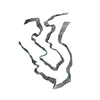
|
|---|---|
| 1 |
|
-Components
| #1: Protein | Mass: 9431.303 Da / Num. of mol.: 6 / Source method: isolated from a natural source / Source: (natural) Homo sapiens (human) / Organ: Heart / Tissue: heart muscle / References: UniProt: P01714*PLUSHas protein modification | Y | |
|---|
-Experimental details
-Experiment
| Experiment | Method: ELECTRON MICROSCOPY |
|---|---|
| EM experiment | Aggregation state: FILAMENT / 3D reconstruction method: helical reconstruction |
- Sample preparation
| Component | Name: Amyloid fibril of an antibody lambda 3 immunoglobulin light chain Type: COMPLEX Details: Extracted fibrils from the explanted heart of a systemic AL amyloidosis patient Entity ID: all / Source: NATURAL |
|---|---|
| Molecular weight | Experimental value: NO |
| Source (natural) | Organism: Homo sapiens (human) / Organ: Heart / Tissue: Heart muscle |
| Buffer solution | pH: 7 |
| Buffer component | Name: Distilled water / Formula: H2O |
| Specimen | Embedding applied: NO / Shadowing applied: NO / Staining applied: NO / Vitrification applied: YES / Details: Sample in pure water, pH not determined |
| Specimen support | Details: 40 mA / Grid material: COPPER / Grid mesh size: 400 divisions/in. / Grid type: C-flat-1.2/1.3 |
| Vitrification | Instrument: FEI VITROBOT MARK III / Cryogen name: ETHANE / Humidity: 95 % / Chamber temperature: 295 K / Details: blot for 9s before plunging |
- Electron microscopy imaging
| Experimental equipment |  Model: Titan Krios / Image courtesy: FEI Company |
|---|---|
| Microscopy | Model: FEI TITAN KRIOS |
| Electron gun | Electron source:  FIELD EMISSION GUN / Accelerating voltage: 300 kV / Illumination mode: FLOOD BEAM FIELD EMISSION GUN / Accelerating voltage: 300 kV / Illumination mode: FLOOD BEAM |
| Electron lens | Mode: BRIGHT FIELD / Cs: 2.7 mm |
| Specimen holder | Cryogen: NITROGEN / Specimen holder model: FEI TITAN KRIOS AUTOGRID HOLDER |
| Image recording | Electron dose: 40 e/Å2 / Detector mode: COUNTING / Film or detector model: GATAN K2 SUMMIT (4k x 4k) / Num. of grids imaged: 1 / Num. of real images: 1964 |
| EM imaging optics | Energyfilter slit width: 20 eV |
| Image scans | Width: 3838 / Height: 3710 / Movie frames/image: 40 |
- Processing
| EM software |
| ||||||||||||||||||||||||||||||
|---|---|---|---|---|---|---|---|---|---|---|---|---|---|---|---|---|---|---|---|---|---|---|---|---|---|---|---|---|---|---|---|
| Image processing | Details: Motion-corrected and dose-weighted movie frames | ||||||||||||||||||||||||||||||
| CTF correction | Details: CTF was estimated from the non-dose-weighted micrographs Type: PHASE FLIPPING AND AMPLITUDE CORRECTION | ||||||||||||||||||||||||||||||
| Helical symmerty | Angular rotation/subunit: -1.1 ° / Axial rise/subunit: 4.8 Å / Axial symmetry: C1 | ||||||||||||||||||||||||||||||
| Particle selection | Num. of particles selected: 194502 Details: manual particle picking helical start-end coordinates | ||||||||||||||||||||||||||||||
| 3D reconstruction | Resolution: 3.2 Å / Resolution method: FSC 0.143 CUT-OFF / Num. of particles: 11003 / Algorithm: FOURIER SPACE / Num. of class averages: 1 / Symmetry type: HELICAL | ||||||||||||||||||||||||||||||
| Atomic model building | B value: 73.24 / Protocol: OTHER / Space: REAL Target criteria: REAL-SPACE (WEIGHTED MAP SUM AT ATOM CENTERS) Details: Secondary structure restraints and NCS were applied during refinement |
