 Movie
Movie Controller


+ Open data

- Basic information
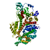
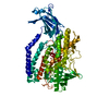
| Entry | Database: PDB / ID: 6ic3 | ||||||
|---|---|---|---|---|---|---|---|
| Title | AL amyloid fibril from a lambda 1 light chain | ||||||
 Components Components | lambda 1 light chain fragment, residues 3-118 | ||||||
 Keywords Keywords | PROTEIN FIBRIL / amyloid fibril / beta sheet / antibody / heart | ||||||
| Biological species |  Homo sapiens (human) Homo sapiens (human) | ||||||
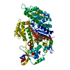
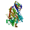
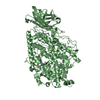
| Method | ELECTRON MICROSCOPY / helical reconstruction / cryo EM / Resolution: 3.3 Å | ||||||
 Authors Authors | Fritz, G. / Faendrich, M. / Schmidt, M. / Radamaker, L. | ||||||
 Citation Citation | Journal: Nat Commun / Year: 2019 Title: Cryo-EM structure of a light chain-derived amyloid fibril from a patient with systemic AL amyloidosis. Authors: Lynn Radamaker / Yin-Hsi Lin / Karthikeyan Annamalai / Stefanie Huhn / Ute Hegenbart / Stefan O Schönland / Günter Fritz / Matthias Schmidt / Marcus Fändrich /  Abstract: Amyloid fibrils derived from antibody light chains are key pathogenic agents in systemic AL amyloidosis. They can be deposited in multiple organs but cardiac amyloid is the major risk factor of ...Amyloid fibrils derived from antibody light chains are key pathogenic agents in systemic AL amyloidosis. They can be deposited in multiple organs but cardiac amyloid is the major risk factor of mortality. Here we report the structure of a λ1 AL amyloid fibril from an explanted human heart at a resolution of 3.3 Å which we determined using cryo-electron microscopy. The fibril core consists of a 91-residue segment presenting an all-beta fold with ten mutagenic changes compared to the germ line. The conformation differs substantially from natively folded light chains: a rotational switch around the intramolecular disulphide bond being the crucial structural rearrangement underlying fibril formation. Our structure provides insight into the mechanism of protein misfolding and the role of patient-specific mutations in pathogenicity. | ||||||
| History |
|
- Structure visualization
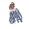
| Movie |
 Movie viewer Movie viewer |
|---|---|
| Structure viewer | Molecule: MolmilJmol/JSmol |
- Downloads & links
-Download
| PDBx/mmCIF format | 6ic3.cif.gz | 128.9 KB | Display | PDBx/mmCIF format |
|---|---|---|---|---|
| PDB format | pdb6ic3.ent.gz | 101.6 KB | Display | PDB format |
| PDBx/mmJSON format | 6ic3.json.gz | Tree view | PDBx/mmJSON format | |
| Others |  Other downloads Other downloads |
-Validation report
| Arichive directory | https://data.pdbj.org/pub/pdb/validation_reports/ic/6ic3ftp://data.pdbj.org/pub/pdb/validation_reports/ic/6ic3 | HTTPS FTP |
|---|
-Related structure data
| Related structure data | 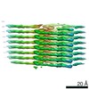 4452MC M: map data used to model this data C: citing same article ( |
|---|---|
| Similar structure data | |
| EM raw data | EMPIAR-10245 (Title: AL amyloid fibril from a lambda 1 light chain / Data size: 44.7 Data #1: Aligned, motion-corrected micrographs of AL fibrils extracted from human heart tissue [micrographs - single frame]) |


-Links
 PDBj
PDBj


- Assembly
| Deposited unit | 
|
|---|---|
| 1 |
|
-Components
| #1: Antibody | Mass: 12177.463 Da / Num. of mol.: 8 / Source method: isolated from a natural source / Source: (natural) Homo sapiens (human) / Organ: Heart / Tissue: heart muscleHas protein modification | Y | |
|---|
-Experimental details
-Experiment
| Experiment | Method: ELECTRON MICROSCOPY |
|---|---|
| EM experiment | Aggregation state: FILAMENT / 3D reconstruction method: helical reconstruction |
- Sample preparation
| Component | Name: Amyloid fibril of an antibody lambda 1 light chain / Type: COMPLEX Details: Extracted fibrils from the heart of a patient suffering from systemic AL amyloidosis Entity ID: all / Source: NATURAL |
|---|---|
| Molecular weight | Value: 2.5 kDa/nm / Experimental value: YES |
| Source (natural) | Organism: Homo sapiens (human) / Organ: heart / Tissue: heart muscle |
| Buffer solution | pH: 7 |
| Buffer component | Name: distilled water / Formula: H2O |
| Specimen | Embedding applied: NO / Shadowing applied: NO / Staining applied: NO / Vitrification applied: YES / Details: Sample in pure water, pH not determined |
| Specimen support | Grid material: COPPER / Grid mesh size: 400 divisions/in. / Grid type: C-flat-1.2/1.3 4C |
| Vitrification | Instrument: GATAN CRYOPLUNGE 3 / Cryogen name: ETHANE / Humidity: 80 % / Chamber temperature: 294 K / Details: blotted from the backside for 4 s before plunging |
- Electron microscopy imaging
| Experimental equipment |  Model: Titan Krios / Image courtesy: FEI Company |
|---|---|
| Microscopy | Model: FEI TITAN KRIOS |
| Electron gun | Electron source:  FIELD EMISSION GUN / Accelerating voltage: 300 kV / Illumination mode: FLOOD BEAM FIELD EMISSION GUN / Accelerating voltage: 300 kV / Illumination mode: FLOOD BEAM |
| Electron lens | Mode: BRIGHT FIELD / Cs: 2.7 mm |
| Specimen holder | Cryogen: NITROGEN / Specimen holder model: FEI TITAN KRIOS AUTOGRID HOLDER |
| Image recording | Average exposure time: 6 sec. / Electron dose: 32 e/Å2 / Detector mode: COUNTING / Film or detector model: GATAN K2 SUMMIT (4k x 4k) / Num. of grids imaged: 1 / Num. of real images: 847 |
| Image scans | Width: 3838 / Height: 3710 / Movie frames/image: 30 / Used frames/image: 1-30 |
- Processing
| Software | Name: PHENIX / Version: 1.14_3260: / Classification: refinement | |||||||||||||||||||||||||||||||||||||||||||||
|---|---|---|---|---|---|---|---|---|---|---|---|---|---|---|---|---|---|---|---|---|---|---|---|---|---|---|---|---|---|---|---|---|---|---|---|---|---|---|---|---|---|---|---|---|---|---|
| EM software |
| |||||||||||||||||||||||||||||||||||||||||||||
| Image processing | Details: Motion-corrected and dose-weighted movie frames | |||||||||||||||||||||||||||||||||||||||||||||
| CTF correction | Details: CTF was estimated from the non-dose-weighted micrographs Type: PHASE FLIPPING AND AMPLITUDE CORRECTION | |||||||||||||||||||||||||||||||||||||||||||||
| Helical symmerty | Angular rotation/subunit: 0.58 ° / Axial rise/subunit: 4.81 Å / Axial symmetry: C1 | |||||||||||||||||||||||||||||||||||||||||||||
| Particle selection | Num. of particles selected: 119395 Details: manual selection. Sigma contrast 3, lowpass filter 20 A | |||||||||||||||||||||||||||||||||||||||||||||
| 3D reconstruction | Resolution: 3.3 Å / Resolution method: FSC 0.143 CUT-OFF / Num. of particles: 32677 / Algorithm: FOURIER SPACE / Num. of class averages: 1 / Symmetry type: HELICAL | |||||||||||||||||||||||||||||||||||||||||||||
| Atomic model building | B value: 111.4 / Protocol: OTHER / Space: REAL Target criteria: REAL-SPACE (WEIGHTED MAP SUM AT ATOM CENTERS) Details: Refinement strategy included global minimization and local grid search and ADP were refined. Secondary structure restraints and NCS were applied during refinement. |
