 Movie
Movie Controller

[English] 日本語
 Yorodumi
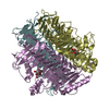
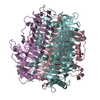
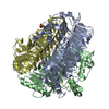
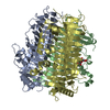
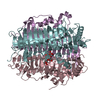
- PDB-5zks: Crystal structure of DFA-IIIase from Arthrobacter chlorophenolicus A6 -
+ Open data

- Basic information
| Entry | Database: PDB / ID: 5zks | ||||||
|---|---|---|---|---|---|---|---|
| Title | Crystal structure of DFA-IIIase from Arthrobacter chlorophenolicus A6 | ||||||
 Components Components | DFA-IIIase | ||||||
 Keywords Keywords | HYDROLASE / hydrolyze difructose anhydride / parallel beta helix / endoenzyme | ||||||
| Function / homology |  Function and homology information Function and homology informationinulin fructotransferase (DFA-I-forming) / inulin fructotransferase (DFA-I-forming) activity / transferase activity Similarity search - Function | ||||||
| Biological species |  Arthrobacter chlorophenolicus A6 (bacteria) Arthrobacter chlorophenolicus A6 (bacteria) | ||||||
| Method |  X-RAY DIFFRACTION / MOLECULAR REPLACEMENT / Resolution: 2 Å X-RAY DIFFRACTION / MOLECULAR REPLACEMENT / Resolution: 2 Å | ||||||
 Authors Authors | Yu, S.H. / Shen, H. / Li, X. / Mu, W.M. | ||||||
 Citation Citation | Journal: Acs Catalysis / Year: 2018 Title: Structural and functional basis of difructose anhydride III hydrolase, which sequentially converts inulin using the same catalytic residue Authors: Yu, S.H. / Shen, H. / Cheng, Y.Y. / Zhu, Y.Y. / Li, X. / Mu, W.M. | ||||||
| History |
|
- Structure visualization
| Structure viewer | Molecule: MolmilJmol/JSmol |
|---|
- Downloads & links
-Download
| PDBx/mmCIF format | 5zks.cif.gz | 182.5 KB | Display | PDBx/mmCIF format |
|---|---|---|---|---|
| PDB format | pdb5zks.ent.gz | 144.7 KB | Display | PDB format |
| PDBx/mmJSON format | 5zks.json.gz | Tree view | PDBx/mmJSON format | |
| Others |  Other downloads Other downloads |
-Validation report
| Arichive directory | https://data.pdbj.org/pub/pdb/validation_reports/zk/5zksftp://data.pdbj.org/pub/pdb/validation_reports/zk/5zks | HTTPS FTP |
|---|
-Related structure data
| Related structure data |  5zkuC  5zkwC  5zkyC  5zl4C  5zl5C  5zlaC  2inuS S: Starting model for refinement C: citing same article ( |
|---|---|
| Similar structure data |
-Links
 PDBj
PDBj- Assembly
| Deposited unit | 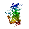
| |||||||||
|---|---|---|---|---|---|---|---|---|---|---|
| 1 | 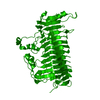
| |||||||||
| Unit cell |
| |||||||||
| Components on special symmetry positions |
|
-Components
| #1: Protein | Mass: 47197.953 Da / Num. of mol.: 1 Source method: isolated from a genetically manipulated source Source: (gene. exp.) Arthrobacter chlorophenolicus A6 (bacteria)Gene: Achl_2895 Production host: References: UniProt: B8HDZ1, Hydrolases; Glycosylases; Glycosidases, i.e. enzymes that hydrolyse O- and S-glycosyl compounds |
|---|---|
| #2: Water | ChemComp-HOH /  Mass: 18.015 Da / Num. of mol.: 207 / Source method: isolated from a natural source / Formula: H2O Mass: 18.015 Da / Num. of mol.: 207 / Source method: isolated from a natural source / Formula: H2O |
-Experimental details
-Experiment
| Experiment | Method: X-RAY DIFFRACTION / Number of used crystals: 1 |
|---|
- Sample preparation
| Crystal | Density Matthews: 4.34 Å3/Da / Density % sol: 71.63 % |
|---|---|
| Crystal grow | Temperature: 287 K / Method: vapor diffusion, hanging drop / Details: 10% PEG 3350, 0.1M Sodium malonate (pH 4.2) |
-Data collection
| Diffraction | Mean temperature: 100 K / Serial crystal experiment: N | |||||||||||||||
|---|---|---|---|---|---|---|---|---|---|---|---|---|---|---|---|---|
| Diffraction source | Source: ROTATING ANODE / Type: RIGAKU / Wavelength: 1.54178 Å | |||||||||||||||
| Detector | Type: RIGAKU RAXIS IV++ / Detector: IMAGE PLATE / Date: Jul 30, 2014 | |||||||||||||||
| Radiation | Protocol: SINGLE WAVELENGTH / Monochromatic (M) / Laue (L): M / Scattering type: x-ray | |||||||||||||||
| Radiation wavelength | Wavelength: 1.54178 Å / Relative weight: 1 | |||||||||||||||
| Reflection twin |
| |||||||||||||||
| Reflection | Resolution: 2→50 Å / Num. obs: 54535 / % possible obs: 99.8 % / Redundancy: 5.4 % / Net I/σ(I): 21.8 | |||||||||||||||
| Reflection shell | Resolution: 2→2.03 Å |
- Processing
| Software |
| ||||||||||||||||||||||||||||||||||||||||||||||||||||||||||||||||||||||||||||||||||||||||||||||||||||||||||||||||||||||||||||||||||||||||||||||||||||||||||||||||||||||||||||||||||||||
|---|---|---|---|---|---|---|---|---|---|---|---|---|---|---|---|---|---|---|---|---|---|---|---|---|---|---|---|---|---|---|---|---|---|---|---|---|---|---|---|---|---|---|---|---|---|---|---|---|---|---|---|---|---|---|---|---|---|---|---|---|---|---|---|---|---|---|---|---|---|---|---|---|---|---|---|---|---|---|---|---|---|---|---|---|---|---|---|---|---|---|---|---|---|---|---|---|---|---|---|---|---|---|---|---|---|---|---|---|---|---|---|---|---|---|---|---|---|---|---|---|---|---|---|---|---|---|---|---|---|---|---|---|---|---|---|---|---|---|---|---|---|---|---|---|---|---|---|---|---|---|---|---|---|---|---|---|---|---|---|---|---|---|---|---|---|---|---|---|---|---|---|---|---|---|---|---|---|---|---|---|---|---|---|
| Refinement | Method to determine structure: MOLECULAR REPLACEMENT Starting model: 2inu Resolution: 2→38.37 Å / Cor.coef. Fo:Fc: 0.982 / Cor.coef. Fo:Fc free: 0.974 / SU B: 2.619 / SU ML: 0.035 / Cross valid method: THROUGHOUT / ESU R: 0.02 / ESU R Free: 0.016 / Stereochemistry target values: MAXIMUM LIKELIHOOD / Details: HYDROGENS HAVE BEEN ADDED IN THE RIDING POSITIONS
| ||||||||||||||||||||||||||||||||||||||||||||||||||||||||||||||||||||||||||||||||||||||||||||||||||||||||||||||||||||||||||||||||||||||||||||||||||||||||||||||||||||||||||||||||||||||
| Solvent computation | Ion probe radii: 0.8 Å / Shrinkage radii: 0.8 Å / VDW probe radii: 1.2 Å / Solvent model: MASK | ||||||||||||||||||||||||||||||||||||||||||||||||||||||||||||||||||||||||||||||||||||||||||||||||||||||||||||||||||||||||||||||||||||||||||||||||||||||||||||||||||||||||||||||||||||||
| Displacement parameters | Biso mean: 29.319 Å2
| ||||||||||||||||||||||||||||||||||||||||||||||||||||||||||||||||||||||||||||||||||||||||||||||||||||||||||||||||||||||||||||||||||||||||||||||||||||||||||||||||||||||||||||||||||||||
| Refinement step | Cycle: 1 / Resolution: 2→38.37 Å
| ||||||||||||||||||||||||||||||||||||||||||||||||||||||||||||||||||||||||||||||||||||||||||||||||||||||||||||||||||||||||||||||||||||||||||||||||||||||||||||||||||||||||||||||||||||||
| Refine LS restraints |
|