 Movie
Movie Controller

[English] 日本語
 Yorodumi
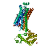
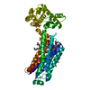
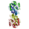
- PDB-6yxh: Cryogenic human alkaline ceramidase 3 (ACER3) at 2.6 A resolution... -
+ Open data

- Basic information
| Entry | Database: PDB / ID: 6yxh | ||||||||||||
|---|---|---|---|---|---|---|---|---|---|---|---|---|---|
| Title | Cryogenic human alkaline ceramidase 3 (ACER3) at 2.6 A resolution determined by Serial Crystallography (SSX) using CrystalDirect | ||||||||||||
 Components Components | Alkaline ceramidase 3 | ||||||||||||
 Keywords Keywords | MEMBRANE PROTEIN / Alkaline ceramidase / ACER3 / serial synchrotron crystallography / SSX / CrystalDirect / LCP crystallisation / in meso / HTX facility / Membrane proteins | ||||||||||||
| Function / homology |  Function and homology information Function and homology informationphytosphingosine biosynthetic process / ceramidase / N-acylsphingosine amidohydrolase activity / ceramide catabolic process / Sphingolipid catabolism / regulation of programmed cell death / sphingosine biosynthetic process / Hydrolases; Acting on carbon-nitrogen bonds, other than peptide bonds; In linear amides / myelination / inflammatory response ...phytosphingosine biosynthetic process / ceramidase / N-acylsphingosine amidohydrolase activity / ceramide catabolic process / Sphingolipid catabolism / regulation of programmed cell death / sphingosine biosynthetic process / Hydrolases; Acting on carbon-nitrogen bonds, other than peptide bonds; In linear amides / myelination / inflammatory response / Golgi membrane / calcium ion binding / positive regulation of cell population proliferation / endoplasmic reticulum membrane / zinc ion binding / membrane Similarity search - Function | ||||||||||||
| Biological species |  Homo sapiens (human) Homo sapiens (human) | ||||||||||||
| Method |  X-RAY DIFFRACTION / SYNCHROTRON / MOLECULAR REPLACEMENT / Resolution: 2.6 Å X-RAY DIFFRACTION / SYNCHROTRON / MOLECULAR REPLACEMENT / Resolution: 2.6 Å | ||||||||||||
 Authors Authors | Healey, R.D. / Basu, S. / Humm, A.S. / Leyrat, C. / Dupeux, F. / Pica, A. / Granier, S. / Marquez, J.A. | ||||||||||||
| Funding support | European Union, 3items
| ||||||||||||
 Citation Citation | Journal: Cell Rep Methods / Year: 2021 Title: An automated platform for structural analysis of membrane proteins through serial crystallography. Authors: Healey, R.D. / Basu, S. / Humm, A.S. / Leyrat, C. / Cong, X. / Golebiowski, J. / Dupeux, F. / Pica, A. / Granier, S. / Marquez, J.A. | ||||||||||||
| History |
|
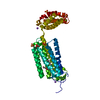
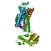
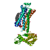
- Structure visualization
| Structure viewer | Molecule: MolmilJmol/JSmol |
|---|
- Downloads & links
-Download
| PDBx/mmCIF format | 6yxh.cif.gz | 160.9 KB | Display | PDBx/mmCIF format |
|---|---|---|---|---|
| PDB format | pdb6yxh.ent.gz | 128.1 KB | Display | PDB format |
| PDBx/mmJSON format | 6yxh.json.gz | Tree view | PDBx/mmJSON format | |
| Others |  Other downloads Other downloads |
-Validation report
| Arichive directory | https://data.pdbj.org/pub/pdb/validation_reports/yx/6yxhftp://data.pdbj.org/pub/pdb/validation_reports/yx/6yxh | HTTPS FTP |
|---|
-Related structure data
| Related structure data |  6yx9C  6yxdC  6yxfC 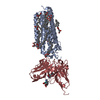 6yxgC  6g7oS S: Starting model for refinement C: citing same article ( |
|---|---|
| Similar structure data |


-Links
 PDBj
PDBj- Assembly
| Deposited unit | 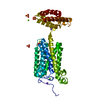
| ||||||||
|---|---|---|---|---|---|---|---|---|---|
| 1 |
| ||||||||
| Unit cell |
|
-Components
-Protein , 1 types, 1 molecules A
| #1: Protein | Mass: 40491.059 Da / Num. of mol.: 1 Source method: isolated from a genetically manipulated source Source: (gene. exp.) Homo sapiens (human) / Gene: ACER3, APHC, PHCA / Production host: Spodoptera frugiperda / Variant (production host): BOLD-2017References: UniProt: Q9NUN7, Hydrolases; Acting on carbon-nitrogen bonds, other than peptide bonds; In linear amides, ceramidase |
|---|
-Non-polymers , 6 types, 72 molecules 


| #2: Chemical | ChemComp-ZN /  Mass: 65.409 Da / Num. of mol.: 1 / Source method: obtained synthetically / Formula: Zn / Feature type: SUBJECT OF INVESTIGATION Mass: 65.409 Da / Num. of mol.: 1 / Source method: obtained synthetically / Formula: Zn / Feature type: SUBJECT OF INVESTIGATION | ||||||||
|---|---|---|---|---|---|---|---|---|---|
| #3: Chemical | ChemComp-SO4 /  Mass: 96.063 Da / Num. of mol.: 6 / Source method: obtained synthetically / Formula: SO4 / Feature type: SUBJECT OF INVESTIGATION Mass: 96.063 Da / Num. of mol.: 6 / Source method: obtained synthetically / Formula: SO4 / Feature type: SUBJECT OF INVESTIGATION#4: Chemical |  Mass: 22.990 Da / Num. of mol.: 2 / Source method: obtained synthetically / Formula: Na / Feature type: SUBJECT OF INVESTIGATION Mass: 22.990 Da / Num. of mol.: 2 / Source method: obtained synthetically / Formula: Na / Feature type: SUBJECT OF INVESTIGATION#5: Chemical | ChemComp-CA / |  Mass: 40.078 Da / Num. of mol.: 1 / Source method: obtained synthetically / Formula: Ca / Feature type: SUBJECT OF INVESTIGATION Mass: 40.078 Da / Num. of mol.: 1 / Source method: obtained synthetically / Formula: Ca / Feature type: SUBJECT OF INVESTIGATION#6: Chemical |  Mass: 24.305 Da / Num. of mol.: 2 / Source method: obtained synthetically / Formula: Mg / Feature type: SUBJECT OF INVESTIGATION Mass: 24.305 Da / Num. of mol.: 2 / Source method: obtained synthetically / Formula: Mg / Feature type: SUBJECT OF INVESTIGATION#7: Water | ChemComp-HOH / | Mass: 18.015 Da / Num. of mol.: 60 / Source method: isolated from a natural source / Formula: H2O |
-Details
| Has ligand of interest | Y |
|---|
-Experimental details
-Experiment
| Experiment | Method: X-RAY DIFFRACTION / Number of used crystals: 1 |
|---|
- Sample preparation
| Crystal | Density Matthews: 3.45 Å3/Da / Density % sol: 64.3 % |
|---|---|
| Crystal grow | Temperature: 293 K / Method: lipidic cubic phase Details: 30 or 50 nl bolus overlaid with 600 nl precipitant solution in CrystalDirect plate-2. 41% PEG 400, 100 mM HEPES pH 7.5, 75 mM magnesium sulfate and 5% DMSO. Crystallisation experiments were ...Details: 30 or 50 nl bolus overlaid with 600 nl precipitant solution in CrystalDirect plate-2. 41% PEG 400, 100 mM HEPES pH 7.5, 75 mM magnesium sulfate and 5% DMSO. Crystallisation experiments were carried out at the HTX facility of EMBL Grenoble. LCP bolus were harvested automatically at cryogenic condition using CrystalDirect technology. |
-Data collection
| Diffraction | Mean temperature: 100 K / Serial crystal experiment: Y |
|---|---|
| Diffraction source | Source: SYNCHROTRON / Site: SLS  / Beamline: X06SA / Wavelength: 0.988 Å / Beamline: X06SA / Wavelength: 0.988 Å |
| Detector | Type: DECTRIS EIGER X 16M / Detector: PIXEL / Date: Feb 23, 2020 |
| Radiation | Protocol: SINGLE WAVELENGTH / Monochromatic (M) / Laue (L): M / Scattering type: x-ray |
| Radiation wavelength | Wavelength: 0.988 Å / Relative weight: 1 |
| Reflection | Resolution: 2.6→50 Å / Num. obs: 17701 / % possible obs: 100 % / Redundancy: 36.01 % / CC1/2: 0.99 / Net I/σ(I): 8.4 |
| Reflection shell | Resolution: 2.6→2.62 Å / Mean I/σ(I) obs: 0.98 / Num. unique obs: 1275 / CC1/2: 0.33 |
| Serial crystallography sample delivery | Method: fixed target |
- Processing
| Software |
| ||||||||||||||||||||||||||||||||||||||||||||||||||||||||||||
|---|---|---|---|---|---|---|---|---|---|---|---|---|---|---|---|---|---|---|---|---|---|---|---|---|---|---|---|---|---|---|---|---|---|---|---|---|---|---|---|---|---|---|---|---|---|---|---|---|---|---|---|---|---|---|---|---|---|---|---|---|---|
| Refinement | Method to determine structure: MOLECULAR REPLACEMENT Starting model: 6G7O Resolution: 2.6→46.18 Å / Cor.coef. Fo:Fc: 0.855 / Cor.coef. Fo:Fc free: 0.839 / SU R Cruickshank DPI: 0.424 / Cross valid method: THROUGHOUT / SU R Blow DPI: 0.428 / SU Rfree Blow DPI: 0.294 / SU Rfree Cruickshank DPI: 0.296
| ||||||||||||||||||||||||||||||||||||||||||||||||||||||||||||
| Displacement parameters | Biso mean: 84.84 Å2
| ||||||||||||||||||||||||||||||||||||||||||||||||||||||||||||
| Refine analyze | Luzzati coordinate error obs: 0.42 Å | ||||||||||||||||||||||||||||||||||||||||||||||||||||||||||||
| Refinement step | Cycle: LAST / Resolution: 2.6→46.18 Å
| ||||||||||||||||||||||||||||||||||||||||||||||||||||||||||||
| Refine LS restraints |
| ||||||||||||||||||||||||||||||||||||||||||||||||||||||||||||
| LS refinement shell | Resolution: 2.6→2.62 Å
| ||||||||||||||||||||||||||||||||||||||||||||||||||||||||||||
| Refinement TLS params. | Origin x: 20.4852 Å / Origin y: 5.6041 Å / Origin z: -40.0276 Å
| ||||||||||||||||||||||||||||||||||||||||||||||||||||||||||||
| Refinement TLS group | Selection details: { A|* } |
