Mass: 18.015 Da / Num. of mol.: 92 / Source method: isolated from a natural source / Formula: H2O
Has protein modification
Y
Sequence details
THIS CONSTRUCT WAS EXPRESSED WITH A PURIFICATION TAG MGSDKIHHHHHHENLYFQG. THE TAG WAS REMOVED WITH ...THIS CONSTRUCT WAS EXPRESSED WITH A PURIFICATION TAG MGSDKIHHHHHHENLYFQG. THE TAG WAS REMOVED WITH TEV PROTEASE LEAVING ONLY A GLYCINE (0) FOLLOWED BY THE TARGET SEQUENCE.
-
Experimental details
-
Experiment
Experiment
Method: X-RAY DIFFRACTION / Number of used crystals: 1
-
Sample preparation
Crystal
Density Matthews: 2.66 Å3/Da / Density % sol: 53.82 %
Crystal grow
Temperature: 293 K / Method: vapor diffusion, sitting drop / pH: 4 Details: 10.0000% polyethylene glycol 6000, 1.0000M lithium chloride, 0.1M citric acid pH 4.0, Additive: 0.001 M Nicotinamide-adenine-dinucleotide (NAD), NANODROP', VAPOR DIFFUSION, SITTING DROP, temperature 293K
Monochromator: Single crystal Si(111) bent monochromator (horizontal focusing) Protocol: MAD / Monochromatic (M) / Laue (L): M / Scattering type: x-ray
Radiation wavelength
ID
Wavelength (Å)
Relative weight
1
0.91837
1
2
0.97947
1
3
0.97883
1
Reflection
Resolution: 2.6→29.527 Å / Num. obs: 48978 / % possible obs: 99.9 % / Redundancy: 4.1 % / Biso Wilson estimate: 58.039 Å2 / Rmerge(I) obs: 0.108 / Rsym value: 0.108 / Net I/σ(I): 11.6
Reflection shell
Diffraction-ID: 1
Resolution (Å)
Redundancy (%)
Rmerge(I) obs
Mean I/σ(I) obs
Num. measured all
Num. unique all
Rsym value
% possible all
2.6-2.67
4.2
0.775
1.9
14957
3564
0.775
100
2.67-2.74
4.2
0.625
1.2
14549
3464
0.625
100
2.74-2.82
4.2
0.543
1.4
14245
3404
0.543
100
2.82-2.91
4.2
0.46
1.6
13870
3308
0.46
100
2.91-3
4.2
0.357
2.1
13429
3206
0.357
100
3-3.11
4.2
0.303
2.5
12929
3087
0.303
100
3.11-3.22
4.2
0.234
3.3
12525
2997
0.234
100
3.22-3.36
4.2
0.172
4.4
12039
2877
0.172
100
3.36-3.51
4.2
0.135
5.5
11541
2762
0.135
100
3.51-3.68
4.2
0.105
6.9
11136
2665
0.105
100
3.68-3.88
4.2
0.091
7.9
10483
2523
0.091
100
3.88-4.11
4.1
0.078
8.8
9955
2399
0.078
100
4.11-4.39
4.1
0.066
10.1
9263
2248
0.066
100
4.39-4.75
4.1
0.055
11.8
8733
2137
0.055
100
4.75-5.2
4.1
0.058
11.5
8027
1959
0.058
100
5.2-5.81
4.1
0.065
10.4
7208
1767
0.065
100
5.81-6.71
4
0.063
10.6
6420
1588
0.063
100
6.71-8.22
4
0.049
12.6
5384
1357
0.049
100
8.22-11.63
3.8
0.041
11.9
4089
1068
0.041
100
11.63-29.53
3.5
0.045
9.5
2093
598
0.045
94.2
-
Phasing
Phasing
Method: MAD
-
Processing
Software
Name
Version
Classification
NB
REFMAC
5.5.0053
refinement
PHENIX
refinement
SOLVE
phasing
MolProbity
3beta29
modelbuilding
SCALA
3.2.5
datascaling
PDB_EXTRACT
3.006
dataextraction
MOSFLM
datareduction
Refinement
Method to determine structure: MAD / Resolution: 2.6→29.527 Å / Cor.coef. Fo:Fc: 0.94 / Cor.coef. Fo:Fc free: 0.916 / Occupancy max: 1 / Occupancy min: 0.5 / SU B: 29.158 / SU ML: 0.269 / TLS residual ADP flag: LIKELY RESIDUAL / Cross valid method: THROUGHOUT / σ(F): 0 / ESU R: 0.613 / ESU R Free: 0.305 Stereochemistry target values: MAXIMUM LIKELIHOOD WITH PHASES Details: 1.HYDROGENS HAVE BEEN ADDED IN THE RIDING POSITIONS. 2.ATOM RECORDS CONTAIN RESIDUAL B FACTORS ONLY. 3.A MET-INHIBITION PROTOCOL WAS USED FOR SELENOMETHIONINE INCORPORATION DURING PROTEIN ...Details: 1.HYDROGENS HAVE BEEN ADDED IN THE RIDING POSITIONS. 2.ATOM RECORDS CONTAIN RESIDUAL B FACTORS ONLY. 3.A MET-INHIBITION PROTOCOL WAS USED FOR SELENOMETHIONINE INCORPORATION DURING PROTEIN EXPRESSION. THE OCCUPANCY OF THE SE ATOMS IN THE MSE RESIDUES WAS REDUCED TO 0.75 FOR THE REDUCED SCATTERING POWER DUE TO PARTIAL S-MET INCORPORATION. 4.A PEG6000 FRAGMENT (PEG) AND 1,2-ETHANEDIOL (EDO) FROM THE CRYSTALLIZATION AND CRYOPROTECTANT SOLUTIONS HAVE BEEN MODELED IN THE SOLVENT STRUCTURE. 5.RAMACHANDRAN OUTLIER AT RESIDUE A294 IS IN A REGION OF POOR ELECTRON DENSITY.
Rfactor
Num. reflection
% reflection
Selection details
Rfree
0.254
2475
5.1 %
RANDOM
Rwork
0.213
-
-
-
obs
0.215
48915
99.9 %
-
Solvent computation
Ion probe radii: 0.8 Å / Shrinkage radii: 0.8 Å / VDW probe radii: 1.4 Å / Solvent model: BABINET MODEL WITH MASK
In the structure databanks used in Yorodumi, some data are registered as the other names, "COVID-19 virus" and "2019-nCoV". Here are the details of the virus and the list of structure data.
Jan 31, 2019. EMDB accession codes are about to change! (news from PDBe EMDB page)
EMDB accession codes are about to change! (news from PDBe EMDB page)
The allocation of 4 digits for EMDB accession codes will soon come to an end. Whilst these codes will remain in use, new EMDB accession codes will include an additional digit and will expand incrementally as the available range of codes is exhausted. The current 4-digit format prefixed with “EMD-” (i.e. EMD-XXXX) will advance to a 5-digit format (i.e. EMD-XXXXX), and so on. It is currently estimated that the 4-digit codes will be depleted around Spring 2019, at which point the 5-digit format will come into force.
The EM Navigator/Yorodumi systems omit the EMD- prefix.
Related info.:Q: What is EMD? / ID/Accession-code notation in Yorodumi/EM Navigator
Yorodumi is a browser for structure data from EMDB, PDB, SASBDB, etc.
This page is also the successor to EM Navigator detail page, and also detail information page/front-end page for Omokage search.
The word "yorodu" (or yorozu) is an old Japanese word meaning "ten thousand". "mi" (miru) is to see.
Related info.:EMDB / PDB / SASBDB / Comparison of 3 databanks / Yorodumi Search / Aug 31, 2016. New EM Navigator & Yorodumi / Yorodumi Papers / Jmol/JSmol / Function and homology information / Changes in new EM Navigator and Yorodumi
 Movie
Movie Controller

 Yorodumi
 Open data
 Basic information
 Components
 Keywords
 Function and homology information
 Corynebacterium glutamicum ATCC 13032 (bacteria)
 X-RAY DIFFRACTION /
  Authors
 Citation
 Structure visualization
 Downloads & links
 Other downloads


 PDBj
PDBj


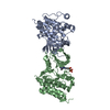
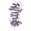
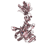
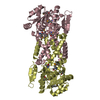
 Assembly


 Mass: 62.068 Da / Num. of mol.: 1 / Source method: obtained synthetically / Formula: C2H6O2
 Mass: 106.120 Da / Num. of mol.: 1 / Source method: obtained synthetically / Formula: C4H10O3
 Mass: 18.015 Da / Num. of mol.: 92 / Source method: isolated from a natural source / Formula: H2O
 Sample preparation
 / Beamline: BL11-1 / Wavelength: 0.91837,0.97947,0.97883
 Processing