- PDB-6g7o: Crystal structure of human alkaline ceramidase 3 (ACER3) at 2.7 A... -
+
Open data
ID or keywords:
Loading...
-
Basic information
Entry
Database: PDB / ID: 6g7o
Title
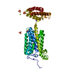
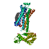
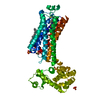
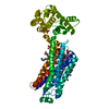
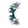
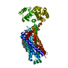
Crystal structure of human alkaline ceramidase 3 (ACER3) at 2.7 Angstrom resolution
Components
Alkaline ceramidase 3,Soluble cytochrome b562
Keywords
MEMBRANE PROTEIN / Hydrolase / 7TM / Zinc binding protein / Calcium-binding protein
Function / homology
Function and homology information
phytosphingosine biosynthetic process / ceramidase / N-acylsphingosine amidohydrolase activity / ceramide catabolic process / Sphingolipid catabolism / regulation of programmed cell death / sphingosine biosynthetic process / Hydrolases; Acting on carbon-nitrogen bonds, other than peptide bonds; In linear amides / myelination / electron transport chain ...phytosphingosine biosynthetic process / ceramidase / N-acylsphingosine amidohydrolase activity / ceramide catabolic process / Sphingolipid catabolism / regulation of programmed cell death / sphingosine biosynthetic process / Hydrolases; Acting on carbon-nitrogen bonds, other than peptide bonds; In linear amides / myelination / electron transport chain / electron transfer activity / periplasmic space / iron ion binding / inflammatory response / Golgi membrane / heme binding / calcium ion binding / positive regulation of cell population proliferation / endoplasmic reticulum membrane / zinc ion binding / membrane Similarity search - Function
In the structure databanks used in Yorodumi, some data are registered as the other names, "COVID-19 virus" and "2019-nCoV". Here are the details of the virus and the list of structure data.
Jan 31, 2019. EMDB accession codes are about to change! (news from PDBe EMDB page)
EMDB accession codes are about to change! (news from PDBe EMDB page)
The allocation of 4 digits for EMDB accession codes will soon come to an end. Whilst these codes will remain in use, new EMDB accession codes will include an additional digit and will expand incrementally as the available range of codes is exhausted. The current 4-digit format prefixed with “EMD-” (i.e. EMD-XXXX) will advance to a 5-digit format (i.e. EMD-XXXXX), and so on. It is currently estimated that the 4-digit codes will be depleted around Spring 2019, at which point the 5-digit format will come into force.
The EM Navigator/Yorodumi systems omit the EMD- prefix.
Related info.:Q: What is EMD? / ID/Accession-code notation in Yorodumi/EM Navigator
Yorodumi is a browser for structure data from EMDB, PDB, SASBDB, etc.
This page is also the successor to EM Navigator detail page, and also detail information page/front-end page for Omokage search.
The word "yorodu" (or yorozu) is an old Japanese word meaning "ten thousand". "mi" (miru) is to see.
Related info.:EMDB / PDB / SASBDB / Comparison of 3 databanks / Yorodumi Search / Aug 31, 2016. New EM Navigator & Yorodumi / Yorodumi Papers / Jmol/JSmol / Function and homology information / Changes in new EM Navigator and Yorodumi
 Movie
Movie Controller

 Yorodumi
 Open data
 Basic information
 Components
 Keywords
 Function and homology information
 Homo sapiens (human)
 X-RAY DIFFRACTION /
  Authors
 France, 1items
  Citation
 Structure visualization
 Downloads & links
 Other downloads
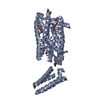


 PDBj
PDBj


 Assembly
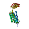

 Spodoptera frugiperda (fall armyworm) / Variant (production host): Sf9


 Mass: 65.409 Da / Num. of mol.: 1 / Source method: obtained synthetically / Formula: Zn
 Mass: 96.063 Da / Num. of mol.: 6 / Source method: obtained synthetically / Formula: SO4
 Mass: 22.990 Da / Num. of mol.: 3 / Source method: obtained synthetically / Formula: Na
 Mass: 356.540 Da / Num. of mol.: 1 / Source method: obtained synthetically / Formula: C21H40O4
 Mass: 40.078 Da / Num. of mol.: 1 / Source method: obtained synthetically / Formula: Ca
 Mass: 24.305 Da / Num. of mol.: 2 / Source method: obtained synthetically / Formula: Mg
 Sample preparation
 / Beamline: X06SA / Wavelength: 1.00001 Å
 Processing