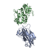
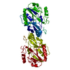
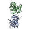
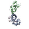
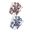
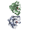
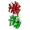
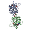
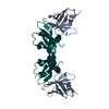
Entry Database : PDB / ID : 5fv2Title Crystal structure of hVEGF in complex with VH domain antibody VASCULAR ENDOTHELIAL GROWTH FACTOR VH DOMAIN ANTIBODY Keywords / / / / Function / homology Function Domain/homology Component
/ / / / / / / / / / / / / / / / / / / / / / / / / / / / / / / / / / / / / / / / / / / / / / / / / / / / / / / / / / / / / / / / / / / / / / / / / / / / / / / / / / / / / / / / / / / / / / / / / / / / / / / / / / / / / / / / / / / / / / / / / / / / / / / / / / / / / / Biological species HOMO SAPIENS (human)Method / / / Resolution : 3.45 Å Authors Chung, C. / Batuwangala, T. Journal : J.Biol.Chem. / Year : 2016Title : Novel Interaction Mechanism of a Domain Antibody Based Inhibitor of Human Vascular Endothelial Growth Factor with Greater Potency Than Ranibizumab and Bevacizumab and Improved Capacity Over Aflibercept.Authors : Walker, A. / Chung, C. / Neu, M. / Burman, M. / Batuwangala, T. / Jones, G. / Tang, C. / Steward, M. / Mullin, M. / Tournier, N. / Lewis, A. / Korczynska, J. / Chung, V. / Catchpole, I. History Deposition Feb 2, 2016 Deposition site / Processing site Revision 1.0 Feb 17, 2016 Provider / Type Revision 1.1 Mar 23, 2016 Group Revision 1.2 May 15, 2019 Group / Experimental preparation / OtherCategory / pdbx_database_proc / pdbx_database_statusItem / _pdbx_database_status.recvd_author_approvalRevision 1.3 Nov 20, 2024 Group Data collection / Database references ... Data collection / Database references / Other / Structure summary Category chem_comp_atom / chem_comp_bond ... chem_comp_atom / chem_comp_bond / database_2 / pdbx_database_status / pdbx_entry_details / pdbx_modification_feature Item _database_2.pdbx_DOI / _database_2.pdbx_database_accession ... _database_2.pdbx_DOI / _database_2.pdbx_database_accession / _pdbx_database_status.status_code_sf / _pdbx_entry_details.has_protein_modification
Show all Show less
 Movie
Movie Controller


 Open data
 Basic information
 Components
 Keywords
 Function and homology information
 HOMO SAPIENS (human)
 X-RAY DIFFRACTION /
  Authors
 Citation
 Structure visualization
 Downloads & links
 Other downloads


 PDBj
PDBj


 Assembly
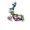


 Sample preparation
 / Type:
  Processing