 Movie
Movie Controller


+ Open data

- Basic information
| Entry | Database: PDB / ID: 6xtb | ||||||
|---|---|---|---|---|---|---|---|
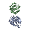
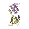
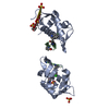
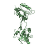
| Title | Subunit BBS 5 of the human core BBSome complex | ||||||
 Components Components | Bardet-Biedl syndrome 5 protein | ||||||
 Keywords Keywords | PROTEIN TRANSPORT / ciliary transport / Arl6 effector / adaptor protein / complex | ||||||
| Function / homology |  Function and homology information Function and homology informationBBSome / motile cilium assembly / BBSome-mediated cargo-targeting to cilium / melanosome transport / phosphatidylinositol-3-phosphate binding / ciliary membrane / heart looping / cilium assembly / axoneme / visual perception ...BBSome / motile cilium assembly / BBSome-mediated cargo-targeting to cilium / melanosome transport / phosphatidylinositol-3-phosphate binding / ciliary membrane / heart looping / cilium assembly / axoneme / visual perception / centriolar satellite / protein transport / RNA polymerase II-specific DNA-binding transcription factor binding / ciliary basal body / cytosol Similarity search - Function | ||||||
| Biological species |  Homo sapiens (human) Homo sapiens (human) | ||||||
| Method | ELECTRON MICROSCOPY / single particle reconstruction / cryo EM / Resolution: 4.3 Å | ||||||
 Authors Authors | Klink, B.U. / Raunser, S. / Gatsogiannis, C. | ||||||
| Funding support |  Germany, 1items Germany, 1items
| ||||||
 Citation Citation | Journal: Elife / Year: 2020 Title: Structure of the human BBSome core complex. Authors: Björn Udo Klink / Christos Gatsogiannis / Oliver Hofnagel / Alfred Wittinghofer / Stefan Raunser / Abstract: The BBSome is a heterooctameric protein complex that plays a central role in primary cilia homeostasis. Its malfunction causes the severe ciliopathy Bardet-Biedl syndrome (BBS). The complex acts as a ...The BBSome is a heterooctameric protein complex that plays a central role in primary cilia homeostasis. Its malfunction causes the severe ciliopathy Bardet-Biedl syndrome (BBS). The complex acts as a cargo adapter that recognizes signaling proteins such as GPCRs and links them to the intraflagellar transport machinery. The underlying mechanism is poorly understood. Here we present a high-resolution cryo-EM structure of a human heterohexameric core subcomplex of the BBSome. The structure reveals the architecture of the complex in atomic detail. It explains how the subunits interact with each other and how disease-causing mutations hamper this interaction. The complex adopts a conformation that is open for binding to membrane-associated GTPase Arl6 and a large positively charged patch likely strengthens the interaction with the membrane. A prominent negatively charged cleft at the center of the complex is likely involved in binding of positively charged signaling sequences of cargo proteins. | ||||||
| History |
|
- Structure visualization
| Movie |
Movie viewer |
|---|---|
| Structure viewer | Molecule: MolmilJmol/JSmol |
- Downloads & links
-Download
| PDBx/mmCIF format | 6xtb.cif.gz | 47.5 KB | Display | PDBx/mmCIF format |
|---|---|---|---|---|
| PDB format | pdb6xtb.ent.gz | 27.9 KB | Display | PDB format |
| PDBx/mmJSON format | 6xtb.json.gz | Tree view | PDBx/mmJSON format | |
| Others |  Other downloads Other downloads |
-Validation report
| Arichive directory | https://data.pdbj.org/pub/pdb/validation_reports/xt/6xtbftp://data.pdbj.org/pub/pdb/validation_reports/xt/6xtb | HTTPS FTP |
|---|
-Related structure data
| Related structure data |  10618MC  6xt9C C: citing same article ( M: map data used to model this data |
|---|---|
| Similar structure data |


-Links
 PDBj
PDBj- Assembly
| Deposited unit | 
|
|---|---|
| 1 |
|
-Components
| #1: Protein | Mass: 38797.926 Da / Num. of mol.: 1 Source method: isolated from a genetically manipulated source Source: (gene. exp.) Homo sapiens (human) / Gene: BBS5 / Cell line (production host): Hi5 / Production host:  Trichoplusia ni (cabbage looper) / References: UniProt: Q8N3I7 Trichoplusia ni (cabbage looper) / References: UniProt: Q8N3I7 |
|---|
-Experimental details
-Experiment
| Experiment | Method: ELECTRON MICROSCOPY |
|---|---|
| EM experiment | Aggregation state: PARTICLE / 3D reconstruction method: single particle reconstruction |
- Sample preparation
| Component | Name: Human BBSome core complex / Type: COMPLEX Details: BBSome core complex containing BBS1,4,5,8,9 and 18. Only BBS5 was modelled into this map since we obtained another map with higher resolution for the other subunits (see related entry) Entity ID: all / Source: RECOMBINANT | ||||||||||||||||
|---|---|---|---|---|---|---|---|---|---|---|---|---|---|---|---|---|---|
| Molecular weight | Experimental value: NO | ||||||||||||||||
| Source (natural) | Organism: Homo sapiens (human) | ||||||||||||||||
| Source (recombinant) | Organism: Trichoplusia ni (cabbage looper) / Strain: Hi5 / Plasmid: ACEMBL | ||||||||||||||||
| Buffer solution | pH: 7.5 | ||||||||||||||||
| Buffer component |
| ||||||||||||||||
| Specimen | Conc.: 0.08 mg/ml / Embedding applied: NO / Shadowing applied: NO / Staining applied: NO / Vitrification applied: YES Details: The sample was cross linked with 0.5% glutaraldehyde | ||||||||||||||||
| Vitrification | Instrument: FEI VITROBOT MARK III / Cryogen name: ETHANE / Humidity: 100 % / Chamber temperature: 286 K Details: double blot with 2 minutes incubation after first sample application |
- Electron microscopy imaging
| Experimental equipment |  Model: Titan Krios / Image courtesy: FEI Company |
|---|---|
| Microscopy | Model: FEI TITAN KRIOS |
| Electron gun | Electron source:  FIELD EMISSION GUN / Accelerating voltage: 300 kV / Illumination mode: OTHER FIELD EMISSION GUN / Accelerating voltage: 300 kV / Illumination mode: OTHER |
| Electron lens | Mode: BRIGHT FIELD / Calibrated defocus min: 300 nm / Calibrated defocus max: 1000 nm / Cs: 2.7 mm |
| Specimen holder | Cryogen: NITROGEN / Specimen holder model: FEI TITAN KRIOS AUTOGRID HOLDER |
| Image recording | Average exposure time: 15 sec. / Electron dose: 67 e/Å2 / Detector mode: COUNTING / Film or detector model: GATAN K2 SUMMIT (4k x 4k) / Num. of real images: 15266 |
| EM imaging optics | Energyfilter slit width: 20 eV / Phase plate: VOLTA PHASE PLATE |
| Image scans | Movie frames/image: 50 / Used frames/image: 1-50 |
- Processing
| EM software |
| |||||||||||||||||||||||||||||||||||||||||||||
|---|---|---|---|---|---|---|---|---|---|---|---|---|---|---|---|---|---|---|---|---|---|---|---|---|---|---|---|---|---|---|---|---|---|---|---|---|---|---|---|---|---|---|---|---|---|---|
| CTF correction | Type: PHASE FLIPPING AND AMPLITUDE CORRECTION | |||||||||||||||||||||||||||||||||||||||||||||
| Particle selection | Num. of particles selected: 2831329 | |||||||||||||||||||||||||||||||||||||||||||||
| Symmetry | Point symmetry: C1 (asymmetric) | |||||||||||||||||||||||||||||||||||||||||||||
| 3D reconstruction | Resolution: 4.3 Å / Resolution method: FSC 0.143 CUT-OFF / Num. of particles: 180654 / Num. of class averages: 1 / Symmetry type: POINT |
