 Movie
Movie Controller

[English] 日本語
 Yorodumi
- PDB-2zfw: Crystal structure of Pex from Synechococcus sp. (strain PCC 7942)... -
+ Open data

- Basic information
| Entry | Database: PDB / ID: 2zfw | ||||||
|---|---|---|---|---|---|---|---|
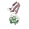
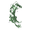
| Title | Crystal structure of Pex from Synechococcus sp. (strain PCC 7942) (Anacystis nidulans R2) | ||||||
 Components Components | Pex | ||||||
 Keywords Keywords | CIRCADIAN CLOCK PROTEIN / Five alpha-helices + One beta-sheet | ||||||
| Function / homology |  Function and homology information Function and homology information: / Transcription regulator PadR, N-terminal / Transcriptional regulator PadR-like family / Winged helix-like DNA-binding domain superfamily/Winged helix DNA-binding domain / Arc Repressor Mutant, subunit A / Winged helix DNA-binding domain superfamily / Winged helix-like DNA-binding domain superfamily / Orthogonal Bundle / Mainly Alpha Similarity search - Domain/homology | ||||||
| Biological species |  Synechococcus sp. (bacteria) Synechococcus sp. (bacteria) | ||||||
| Method |  X-RAY DIFFRACTION / SYNCHROTRON / MIR / Resolution: 2.9 Å X-RAY DIFFRACTION / SYNCHROTRON / MIR / Resolution: 2.9 Å | ||||||
 Authors Authors | Kouyama, T. | ||||||
 Citation Citation | Journal: Genes Cells / Year: 2009 Title: Functionally important structural elements of the cyanobacterial clock-related protein Pex. Authors: Kurosawa, S. / Murakami, R. / Onai, K. / Morishita, M. / Hasegawa, D. / Iwase, R. / Uzumaki, T. / Hayashi, F. / Kitajima-Ihara, T. / Sakata, S. / Murakami, M. / Kouyama, T. / Ishiura, M. | ||||||
| History |
|
- Structure visualization
| Structure viewer | Molecule: MolmilJmol/JSmol |
|---|
- Downloads & links
-Download
| PDBx/mmCIF format | 2zfw.cif.gz | 106 KB | Display | PDBx/mmCIF format |
|---|---|---|---|---|
| PDB format | pdb2zfw.ent.gz | 82.6 KB | Display | PDB format |
| PDBx/mmJSON format | 2zfw.json.gz | Tree view | PDBx/mmJSON format | |
| Others |  Other downloads Other downloads |
-Validation report
| Arichive directory | https://data.pdbj.org/pub/pdb/validation_reports/zf/2zfwftp://data.pdbj.org/pub/pdb/validation_reports/zf/2zfw | HTTPS FTP |
|---|
-Related structure data
| Related structure data | 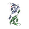 2dqlC 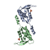 2e1nS C: citing same article ( S: Starting model for refinement |
|---|---|
| Similar structure data |


-Links
 PDBj
PDBj
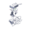
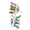
- Assembly
| Deposited unit | 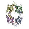
| ||||||||
|---|---|---|---|---|---|---|---|---|---|
| 1 | 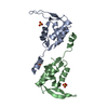
| ||||||||
| 2 | 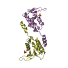
| ||||||||
| Unit cell |
|
-Components
| #1: Protein | Mass: 17162.320 Da / Num. of mol.: 4 Source method: isolated from a genetically manipulated source Source: (gene. exp.) Synechococcus sp. (bacteria) / Strain: PCC 7942 / Gene: pex / Plasmid: pGEX-6P-1 / Production host: #2: Chemical | ChemComp-SO4 /   Mass: 96.063 Da / Num. of mol.: 5 / Source method: obtained synthetically / Formula: SO4 Mass: 96.063 Da / Num. of mol.: 5 / Source method: obtained synthetically / Formula: SO4#3: Water | ChemComp-HOH / |  Mass: 18.015 Da / Num. of mol.: 9 / Source method: isolated from a natural source / Formula: H2O Mass: 18.015 Da / Num. of mol.: 9 / Source method: isolated from a natural source / Formula: H2O |
|---|
-Experimental details
-Experiment
| Experiment | Method: X-RAY DIFFRACTION |
|---|
- Sample preparation
| Crystal | Density Matthews: 2.7 Å3/Da / Density % sol: 54.42 % |
|---|---|
| Crystal grow | Temperature: 283 K / Method: vapor diffusion, hanging drop / pH: 8.5 Details: 2.5M ammonium sulfate, 0.2M Lithium sulfate, 0.1M Tris-HCl, pH 8.5, VAPOR DIFFUSION, HANGING DROP, temperature 283K |
-Data collection
| Diffraction source | Source: SYNCHROTRON / Site: SPring-8  / Beamline: BL41XU / Wavelength: 1 Å / Beamline: BL41XU / Wavelength: 1 Å |
|---|---|
| Detector | Type: ADSC QUANTUM 315 / Detector: CCD / Date: Nov 22, 2006 / Details: mirrors |
| Radiation | Monochromator: double-crystal monochromator / Protocol: SINGLE WAVELENGTH / Monochromatic (M) / Laue (L): M / Scattering type: x-ray |
| Radiation wavelength | Wavelength: 1 Å / Relative weight: 1 |
| Reflection | Resolution: 2.9→100 Å / Num. all: 16173 / Num. obs: 14969 / % possible obs: 100 % / Observed criterion σ(F): 0 / Observed criterion σ(I): 0 / Redundancy: 12.1 % / Biso Wilson estimate: 21.96 Å2 / Rmerge(I) obs: 0.098 / Rsym value: 0.098 / Net I/σ(I): 19 |
| Reflection shell | Resolution: 2.9→3.06 Å / Redundancy: 12.9 % / Rmerge(I) obs: 0.497 / Mean I/σ(I) obs: 3.7 / Num. unique all: 1260 / Rsym value: 0.497 / % possible all: 100 |
- Processing
| Software |
| ||||||||||||||||||||
|---|---|---|---|---|---|---|---|---|---|---|---|---|---|---|---|---|---|---|---|---|---|
| Refinement | Method to determine structure: MIR Starting model: PDB ENTRY 2E1N Resolution: 2.9→15 Å / Isotropic thermal model: Isotropic / Cross valid method: THROUGHOUT / σ(F): 0 / σ(I): 0 / Stereochemistry target values: Engh & Huber
| ||||||||||||||||||||
| Displacement parameters | Biso mean: 71.2 Å2
| ||||||||||||||||||||
| Refine analyze |
| ||||||||||||||||||||
| Refinement step | Cycle: LAST / Resolution: 2.9→15 Å
| ||||||||||||||||||||
| Refine LS restraints |
| ||||||||||||||||||||
| LS refinement shell | Resolution: 2.9→3 Å
|
