 Movie
Movie Controller


+ Open data

- Basic information
| Entry | Database: PDB / ID: 1dyk | ||||||
|---|---|---|---|---|---|---|---|
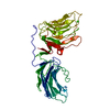
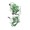
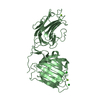
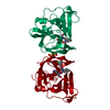
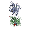
| Title | Laminin alpha 2 chain LG4-5 domain pair | ||||||
 Components Components | LAMININ ALPHA 2 CHAIN | ||||||
 Keywords Keywords | METAL BINDING PROTEIN / LAMININ | ||||||
| Function / homology |  Function and homology information Function and homology informationregulation of basement membrane organization / Schwann cell differentiation / positive regulation of synaptic transmission, cholinergic / positive regulation of integrin-mediated signaling pathway / tissue development / protein complex involved in cell-matrix adhesion / positive regulation of muscle cell differentiation / extracellular matrix structural constituent / basement membrane / regulation of embryonic development ...regulation of basement membrane organization / Schwann cell differentiation / positive regulation of synaptic transmission, cholinergic / positive regulation of integrin-mediated signaling pathway / tissue development / protein complex involved in cell-matrix adhesion / positive regulation of muscle cell differentiation / extracellular matrix structural constituent / basement membrane / regulation of embryonic development / synaptic cleft / positive regulation of cell adhesion / regulation of cell migration / axon guidance / animal organ morphogenesis / neuromuscular junction / sarcolemma / extracellular matrix / dendritic spine / cell adhesion / signaling receptor binding / extracellular region Similarity search - Function | ||||||
| Biological species |  | ||||||
| Method |  X-RAY DIFFRACTION / SYNCHROTRON / MOLECULAR REPLACEMENT / Resolution: 2 Å X-RAY DIFFRACTION / SYNCHROTRON / MOLECULAR REPLACEMENT / Resolution: 2 Å | ||||||
 Authors Authors | Tisi, D. / Talts, J.F. / Timple, R. / Hohenester, E. | ||||||
 Citation Citation | Journal: Embo J. / Year: 2000 Title: Structure of the C-Terminal Laminin G-Like Domain Pair of the Laminin Alpha 2 Chain Harbouring Binding Sites for Alpha-Dystroglycan and Heparin Authors: Tisi, D. / Talts, J.F. / Timpl, R. / Hohenester, E. #1: Journal: Mol.Cell / Year: 1999Title: The Crystal Structure of a Laminin G-Like Module Reveals the Molecular Basis of Alpha-Dystroglycan Binding to Laminins, Perlecan and Agrin Authors: Hohenester, E. / Tisi, D. / Talts, J.F. / Timpl, R. #2: Journal: FEBS Lett. / Year: 1998 Title: Structural Analysis and Proteolytic Processing of Recombinant G Domain of Mouse Laminin Alpha 2 Chain Authors: Talts, J.F. / Mann, K. / Yamada, Y. / Timpl, R. | ||||||
| History |
|
- Structure visualization
| Structure viewer | Molecule: MolmilJmol/JSmol |
|---|
- Downloads & links
-Download
| PDBx/mmCIF format | 1dyk.cif.gz | 91.7 KB | Display | PDBx/mmCIF format |
|---|---|---|---|---|
| PDB format | pdb1dyk.ent.gz | 68.2 KB | Display | PDB format |
| PDBx/mmJSON format | 1dyk.json.gz | Tree view | PDBx/mmJSON format | |
| Others |  Other downloads Other downloads |
-Validation report
| Arichive directory | https://data.pdbj.org/pub/pdb/validation_reports/dy/1dykftp://data.pdbj.org/pub/pdb/validation_reports/dy/1dyk | HTTPS FTP |
|---|
-Related structure data
| Related structure data |  1qu0S S: Starting model for refinement |
|---|---|
| Similar structure data |


-Links
 PDBj
PDBj


- Assembly
| Deposited unit | 
| ||||||||
|---|---|---|---|---|---|---|---|---|---|
| 1 |
| ||||||||
| Unit cell |
|
-Components
| #1: Protein | Mass: 42743.570 Da / Num. of mol.: 1 / Fragment: LAMININ G-LIKE DOMAIN 4-5 PAIR / Mutation: YES Source method: isolated from a genetically manipulated source Source: (gene. exp.)  HOMO SAPIENS (human) / References: UniProt: Q60675 HOMO SAPIENS (human) / References: UniProt: Q60675 | ||||||
|---|---|---|---|---|---|---|---|
| #2: Chemical |   Mass: 40.078 Da / Num. of mol.: 2 / Source method: obtained synthetically / Formula: Ca Mass: 40.078 Da / Num. of mol.: 2 / Source method: obtained synthetically / Formula: Ca#3: Water | ChemComp-HOH / |  Mass: 18.015 Da / Num. of mol.: 251 / Source method: isolated from a natural source / Formula: H2O Mass: 18.015 Da / Num. of mol.: 251 / Source method: isolated from a natural source / Formula: H2OHas protein modification | Y | Sequence details | THE SWISSPROT SEQUENCE IS TAKEN FROM BERNIER S.M., UTANI A., SUGIYAMA S., DOI T., POLISTINA C., ...THE SWISSPROT SEQUENCE IS TAKEN FROM BERNIER S.M., UTANI A., SUGIYAMA S., DOI T., POLISTINA C., YAMADA Y. MATRIX BIOL. 14:447-455(1995). THE 21 RESIDUES OF THE C-TERMINUS DO NOT MATCH WITH THE SEQUENCE DATABASE REFERENCE PROVIDED. THE COORDINATE | |
-Experimental details
-Experiment
| Experiment | Method: X-RAY DIFFRACTION / Number of used crystals: 1 |
|---|
- Sample preparation
| Crystal | Density Matthews: 2.87 Å3/Da / Density % sol: 57.19 % Description: MOLECULAR REPLACEMENT PLUS SIR FROM SM DERIVATIVE | ||||||||||||||||||||||||||||||||||||||||||||||||
|---|---|---|---|---|---|---|---|---|---|---|---|---|---|---|---|---|---|---|---|---|---|---|---|---|---|---|---|---|---|---|---|---|---|---|---|---|---|---|---|---|---|---|---|---|---|---|---|---|---|
| Crystal grow | pH: 7.5 / Details: pH 7.50 | ||||||||||||||||||||||||||||||||||||||||||||||||
| Crystal | *PLUS Density % sol: 60 % | ||||||||||||||||||||||||||||||||||||||||||||||||
| Crystal grow | *PLUS pH: 8 / Method: vapor diffusion, hanging drop | ||||||||||||||||||||||||||||||||||||||||||||||||
| Components of the solutions | *PLUS
|
-Data collection
| Diffraction | Mean temperature: 100 K |
|---|---|
| Diffraction source | Source: SYNCHROTRON / Site: SRS  / Beamline: PX9.6 / Wavelength: 0.87 / Beamline: PX9.6 / Wavelength: 0.87 |
| Detector | Type: ADSC QUANTUM 4 CCD / Detector: CCD / Date: Oct 15, 1999 |
| Radiation | Protocol: SINGLE WAVELENGTH / Monochromatic (M) / Laue (L): M / Scattering type: x-ray |
| Radiation wavelength | Wavelength: 0.87 Å / Relative weight: 1 |
| Reflection | Resolution: 2→20 Å / Num. obs: 31704 / % possible obs: 94.8 % / Redundancy: 3.4 % / Rmerge(I) obs: 0.056 |
| Reflection shell | Resolution: 2→2.11 Å / Redundancy: 3.2 % / Rmerge(I) obs: 0.139 / % possible all: 93.6 |
| Reflection shell | *PLUS % possible obs: 93.6 % |
- Processing
| Software |
| ||||||||||||||||||||||||||||||||||||||||||||||||||||||||||||||||||||||||||||||||
|---|---|---|---|---|---|---|---|---|---|---|---|---|---|---|---|---|---|---|---|---|---|---|---|---|---|---|---|---|---|---|---|---|---|---|---|---|---|---|---|---|---|---|---|---|---|---|---|---|---|---|---|---|---|---|---|---|---|---|---|---|---|---|---|---|---|---|---|---|---|---|---|---|---|---|---|---|---|---|---|---|---|
| Refinement | Method to determine structure: MOLECULAR REPLACEMENT Starting model: 1QU0 Resolution: 2→20 Å / Isotropic thermal model: INDIVIDUAL RESTRAINED / Cross valid method: THROUGHOUT / σ(F): 0
| ||||||||||||||||||||||||||||||||||||||||||||||||||||||||||||||||||||||||||||||||
| Refinement step | Cycle: LAST / Resolution: 2→20 Å
| ||||||||||||||||||||||||||||||||||||||||||||||||||||||||||||||||||||||||||||||||
| Refine LS restraints |
| ||||||||||||||||||||||||||||||||||||||||||||||||||||||||||||||||||||||||||||||||
| Software | *PLUS Name: X-PLOR / Classification: refinement | ||||||||||||||||||||||||||||||||||||||||||||||||||||||||||||||||||||||||||||||||
| Refinement | *PLUS Num. reflection Rfree: 2244 | ||||||||||||||||||||||||||||||||||||||||||||||||||||||||||||||||||||||||||||||||
| Solvent computation | *PLUS | ||||||||||||||||||||||||||||||||||||||||||||||||||||||||||||||||||||||||||||||||
| Displacement parameters | *PLUS |
