 Movie
Movie Controller

[English] 日本語
 Yorodumi
- PDB-1qu0: CRYSTAL STRUCTURE OF THE FIFTH LAMININ G-LIKE MODULE OF THE MOUSE... -
+ Open data

- Basic information
| Entry | Database: PDB / ID: 1qu0 | ||||||
|---|---|---|---|---|---|---|---|
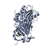
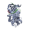
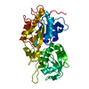
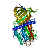
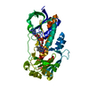
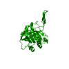
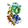
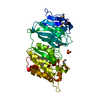
| Title | CRYSTAL STRUCTURE OF THE FIFTH LAMININ G-LIKE MODULE OF THE MOUSE LAMININ ALPHA2 CHAIN | ||||||
 Components Components | LAMININ ALPHA2 CHAIN | ||||||
 Keywords Keywords | METAL BINDING PROTEIN / BETA SANDWICH / CALCIUM-BINDING PROTEIN | ||||||
| Function / homology |  Function and homology information Function and homology informationregulation of basement membrane organization / Schwann cell differentiation / positive regulation of synaptic transmission, cholinergic / positive regulation of integrin-mediated signaling pathway / tissue development / protein complex involved in cell-matrix adhesion / positive regulation of muscle cell differentiation / extracellular matrix structural constituent / basement membrane / regulation of embryonic development ...regulation of basement membrane organization / Schwann cell differentiation / positive regulation of synaptic transmission, cholinergic / positive regulation of integrin-mediated signaling pathway / tissue development / protein complex involved in cell-matrix adhesion / positive regulation of muscle cell differentiation / extracellular matrix structural constituent / basement membrane / regulation of embryonic development / synaptic cleft / regulation of cell migration / positive regulation of cell adhesion / axon guidance / animal organ morphogenesis / neuromuscular junction / sarcolemma / extracellular matrix / dendritic spine / cell adhesion / signaling receptor binding / extracellular region Similarity search - Function | ||||||
| Biological species |  | ||||||
| Method |  X-RAY DIFFRACTION / SYNCHROTRON / Resolution: 2.35 Å X-RAY DIFFRACTION / SYNCHROTRON / Resolution: 2.35 Å | ||||||
 Authors Authors | Hohenester, E. / Tisi, D. / Talts, J.F. / Timpl, R. | ||||||
 Citation Citation | Journal: Mol.Cell / Year: 1999 Title: The crystal structure of a laminin G-like module reveals the molecular basis of alpha-dystroglycan binding to laminins, perlecan, and agrin. Authors: Hohenester, E. / Tisi, D. / Talts, J.F. / Timpl, R. #1: Journal: FEBS Lett. / Year: 1998Title: Structural analysis and proteolytic processing of recombinant G domain of mouse laminin alpha2 chain Authors: Talts, J.F. / Mann, K. / Yamada, Y. / Timpl, R. #2: Journal: J.Anat. / Year: 1998Title: The role of laminins in basement membrane assembly Authors: Aumailley, M. / Smyth, N. | ||||||
| History |
|
- Structure visualization
| Structure viewer | Molecule: MolmilJmol/JSmol |
|---|
- Downloads & links
-Download
| PDBx/mmCIF format | 1qu0.cif.gz | 153.5 KB | Display | PDBx/mmCIF format |
|---|---|---|---|---|
| PDB format | pdb1qu0.ent.gz | 120.8 KB | Display | PDB format |
| PDBx/mmJSON format | 1qu0.json.gz | Tree view | PDBx/mmJSON format | |
| Others |  Other downloads Other downloads |
-Validation report
| Arichive directory | https://data.pdbj.org/pub/pdb/validation_reports/qu/1qu0ftp://data.pdbj.org/pub/pdb/validation_reports/qu/1qu0 | HTTPS FTP |
|---|
-Related structure data
| Similar structure data |
|---|


-Links
 PDBj
PDBj


- Assembly
| Deposited unit | 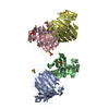
| ||||||||
|---|---|---|---|---|---|---|---|---|---|
| 1 | 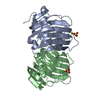
| ||||||||
| 2 | 
| ||||||||
| 3 | 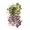
| ||||||||
| 4 | 
| ||||||||
| Unit cell |
| ||||||||
| Details | the asymmetric unit contains four copies of the laminin alpha2 LG5 module, which is monomeric in solution |
-Components
| #1: Protein | Mass: 20420.248 Da / Num. of mol.: 4 / Fragment: LG5 MODULE Mutation: THE N-TERMINAL APLA SEQUENCE IS DERIVED FROM THE VECTOR Source method: isolated from a genetically manipulated source Source: (gene. exp.) #2: Chemical | ChemComp-CA /   Mass: 40.078 Da / Num. of mol.: 4 / Source method: obtained synthetically / Formula: Ca Mass: 40.078 Da / Num. of mol.: 4 / Source method: obtained synthetically / Formula: Ca#3: Chemical | ChemComp-SO4 /   Mass: 96.063 Da / Num. of mol.: 7 / Source method: obtained synthetically / Formula: SO4 Mass: 96.063 Da / Num. of mol.: 7 / Source method: obtained synthetically / Formula: SO4#4: Water | ChemComp-HOH / |  Mass: 18.015 Da / Num. of mol.: 275 / Source method: isolated from a natural source / Formula: H2O Mass: 18.015 Da / Num. of mol.: 275 / Source method: isolated from a natural source / Formula: H2OHas protein modification | Y | |
|---|
-Experimental details
-Experiment
| Experiment | Method: X-RAY DIFFRACTION / Number of used crystals: 1 |
|---|
- Sample preparation
| Crystal | Density Matthews: 3.42 Å3/Da / Density % sol: 64.07 % | ||||||||||||||||||||||||||||||||||||||||||
|---|---|---|---|---|---|---|---|---|---|---|---|---|---|---|---|---|---|---|---|---|---|---|---|---|---|---|---|---|---|---|---|---|---|---|---|---|---|---|---|---|---|---|---|
| Crystal grow | Temperature: 291 K / Method: vapor diffusion, hanging drop / pH: 7.5 Details: 2 M ammonium sulfate, 2% isopropanol, 100 mM HEPES, 10 mM calcium chloride, pH 7.5, VAPOR DIFFUSION, HANGING DROP, temperature 291K | ||||||||||||||||||||||||||||||||||||||||||
| Crystal grow | *PLUS Temperature: 19 ℃Details: drop consists of equal volume of protein and reservoir solutions | ||||||||||||||||||||||||||||||||||||||||||
| Components of the solutions | *PLUS
|
-Data collection
| Diffraction | Mean temperature: 100 K |
|---|---|
| Diffraction source | Source: SYNCHROTRON / Site: SRS  / Beamline: PX7.2 / Wavelength: 1.488 / Beamline: PX7.2 / Wavelength: 1.488 |
| Detector | Type: MARRESEARCH / Detector: IMAGE PLATE / Date: Feb 19, 1999 |
| Radiation | Protocol: SINGLE WAVELENGTH / Monochromatic (M) / Laue (L): M / Scattering type: x-ray |
| Radiation wavelength | Wavelength: 1.488 Å / Relative weight: 1 |
| Reflection | Resolution: 2.35→20 Å / Num. all: 43258 / Num. obs: 43258 / % possible obs: 94.3 % / Observed criterion σ(I): 0 / Redundancy: 2.1 % / Biso Wilson estimate: 28 Å2 / Rmerge(I) obs: 0.053 / Net I/σ(I): 12.6 |
| Reflection shell | Resolution: 2.35→2.48 Å / Redundancy: 2 % / Rmerge(I) obs: 0.11 / % possible all: 85.8 |
| Reflection shell | *PLUS % possible obs: 85.8 % |
- Processing
| Software |
| ||||||||||||||||||||
|---|---|---|---|---|---|---|---|---|---|---|---|---|---|---|---|---|---|---|---|---|---|
| Refinement | Resolution: 2.35→20 Å / σ(F): 0 / Stereochemistry target values: Engh & Huber / Details: bulk solvent correction applied
| ||||||||||||||||||||
| Refinement step | Cycle: LAST / Resolution: 2.35→20 Å
| ||||||||||||||||||||
| Refine LS restraints |
| ||||||||||||||||||||
| Software | *PLUS Name: X-PLOR / Version: 3.851 / Classification: refinement | ||||||||||||||||||||
| Refinement | *PLUS Num. reflection obs: 41083 | ||||||||||||||||||||
| Solvent computation | *PLUS | ||||||||||||||||||||
| Displacement parameters | *PLUS |
