 Movie
Movie Controller


+ Open data

- Basic information
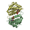
| Entry | Database: PDB / ID: 5cdx | ||||||
|---|---|---|---|---|---|---|---|
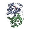
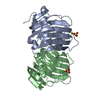
| Title | Crystal structure of conserpin | ||||||
 Components Components | Conserpin | ||||||
 Keywords Keywords | hydrolase inhibitor / Serine Protease Inhibitor / Aggregation Resistant / Consensus Design / Stability | ||||||
| Function / homology | Antithrombin; Chain I, domain 2 / Antithrombin, subunit I, domain 2 / Alpha-1-antitrypsin; domain 1 / Alpha-1-antitrypsin, domain 1 / Roll / 2-Layer Sandwich / Mainly Beta / Alpha Beta Function and homology information Function and homology information | ||||||
| Biological species | synthetic construct (others) | ||||||
| Method |  X-RAY DIFFRACTION / SYNCHROTRON / MOLECULAR REPLACEMENT / Resolution: 2.4 Å X-RAY DIFFRACTION / SYNCHROTRON / MOLECULAR REPLACEMENT / Resolution: 2.4 Å | ||||||
 Authors Authors | Porebski, B.T. / Borg, N.A. / McGowan, S. / Buckle, A.M. | ||||||
 Citation Citation | Journal: Sci Rep / Year: 2016 Title: Smoothing a rugged protein folding landscape by sequence-based redesign. Authors: Porebski, B.T. / Keleher, S. / Hollins, J.J. / Nickson, A.A. / Marijanovic, E.M. / Borg, N.A. / Costa, M.G. / Pearce, M.A. / Dai, W. / Zhu, L. / Irving, J.A. / Hoke, D.E. / Kass, I. / ...Authors: Porebski, B.T. / Keleher, S. / Hollins, J.J. / Nickson, A.A. / Marijanovic, E.M. / Borg, N.A. / Costa, M.G. / Pearce, M.A. / Dai, W. / Zhu, L. / Irving, J.A. / Hoke, D.E. / Kass, I. / Whisstock, J.C. / Bottomley, S.P. / Webb, G.I. / McGowan, S. / Buckle, A.M. | ||||||
| History |
|
- Structure visualization
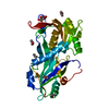
| Structure viewer | Molecule: MolmilJmol/JSmol |
|---|
- Downloads & links
-Download
| PDBx/mmCIF format | 5cdx.cif.gz | 148.7 KB | Display | PDBx/mmCIF format |
|---|---|---|---|---|
| PDB format | pdb5cdx.ent.gz | 115.5 KB | Display | PDB format |
| PDBx/mmJSON format | 5cdx.json.gz | Tree view | PDBx/mmJSON format | |
| Others |  Other downloads Other downloads |
-Validation report
| Arichive directory | https://data.pdbj.org/pub/pdb/validation_reports/cd/5cdxftp://data.pdbj.org/pub/pdb/validation_reports/cd/5cdx | HTTPS FTP |
|---|
-Related structure data
| Related structure data | 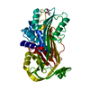 5cdzC 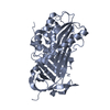 5ce0C  3ne4S  5ce2 C: citing same article ( S: Starting model for refinement |
|---|---|
| Similar structure data |


-Links
 PDBj
PDBj- Assembly
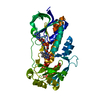
| Deposited unit | 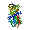
| ||||||||
|---|---|---|---|---|---|---|---|---|---|
| 1 |
| ||||||||
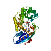
| Unit cell |
|
-Components
| #1: Protein | Mass: 42578.332 Da / Num. of mol.: 1 Source method: isolated from a genetically manipulated source Source: (gene. exp.) synthetic construct (others) / Production host:  |
|---|---|
| #2: Water | ChemComp-HOH /  Mass: 18.015 Da / Num. of mol.: 58 / Source method: isolated from a natural source / Formula: H2O Mass: 18.015 Da / Num. of mol.: 58 / Source method: isolated from a natural source / Formula: H2O |
-Experimental details
-Experiment
| Experiment | Method: X-RAY DIFFRACTION |
|---|
- Sample preparation
| Crystal | Density Matthews: 2.29 Å3/Da / Density % sol: 46.24 % |
|---|---|
| Crystal grow | Temperature: 288 K / Method: vapor diffusion, hanging drop / pH: 7.5 Details: Crystals appeared within 5 days in 0.2 M magnesium chloride hexahydrate, 16% PEG-3350, 10 mM bis-tris, pH 7.5 |
-Data collection
| Diffraction | Mean temperature: 100 K |
|---|---|
| Diffraction source | Source: SYNCHROTRON / Site: Australian Synchrotron  / Beamline: MX2 / Wavelength: 0.95369 Å / Beamline: MX2 / Wavelength: 0.95369 Å |
| Detector | Type: ADSC QUANTUM 315r / Detector: CCD / Date: Dec 16, 2010 |
| Radiation | Monochromator: Silicon Double Crystal / Protocol: SINGLE WAVELENGTH / Monochromatic (M) / Laue (L): M / Scattering type: x-ray |
| Radiation wavelength | Wavelength: 0.95369 Å / Relative weight: 1 |
| Reflection | Resolution: 2.4→42.06 Å / Num. all: 30073 / Num. obs: 15325 / % possible obs: 97.77 % / Redundancy: 2 % / Rmerge(I) obs: 0.04451 / Net I/σ(I): 9.2 |
| Reflection shell | Resolution: 2.4→2.486 Å / Redundancy: 2 % / Mean I/σ(I) obs: 2.16 / Num. unique all: 1498 / % possible all: 97.46 |
- Processing
| Software |
| ||||||||||||||||||||||||||||||||||||||||||
|---|---|---|---|---|---|---|---|---|---|---|---|---|---|---|---|---|---|---|---|---|---|---|---|---|---|---|---|---|---|---|---|---|---|---|---|---|---|---|---|---|---|---|---|
| Refinement | Method to determine structure: MOLECULAR REPLACEMENT Starting model: 3NE4 Resolution: 2.4→42.06 Å / SU ML: 0.39 / Cross valid method: FREE R-VALUE / σ(F): 1.34 / Phase error: 32.35 / Stereochemistry target values: ML
| ||||||||||||||||||||||||||||||||||||||||||
| Solvent computation | Shrinkage radii: 0.98 Å / VDW probe radii: 1.2 Å / Solvent model: FLAT BULK SOLVENT MODEL / Bsol: 40.917 Å2 / ksol: 0.315 e/Å3 | ||||||||||||||||||||||||||||||||||||||||||
| Displacement parameters |
| ||||||||||||||||||||||||||||||||||||||||||
| Refinement step | Cycle: LAST / Resolution: 2.4→42.06 Å
| ||||||||||||||||||||||||||||||||||||||||||
| Refine LS restraints |
| ||||||||||||||||||||||||||||||||||||||||||
| LS refinement shell |
| ||||||||||||||||||||||||||||||||||||||||||
| Refinement TLS params. | Method: refined / Origin x: 36.2747 Å / Origin y: 44.5292 Å / Origin z: 57.1267 Å
| ||||||||||||||||||||||||||||||||||||||||||
| Refinement TLS group | Selection details: all |
