 Movie
Movie Controller

[English] 日本語
 Yorodumi
- PDB-3guz: Structural and substrate-binding studies of pantothenate synthena... -
+ Open data

- Basic information
| Entry | Database: PDB / ID: 3guz | ||||||
|---|---|---|---|---|---|---|---|
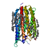
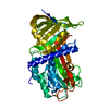
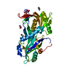
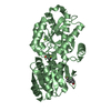
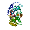
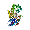
| Title | Structural and substrate-binding studies of pantothenate synthenate (PS)provide insights into homotropic inhibition by pantoate in PS's | ||||||
 Components Components | Pantothenate synthetase | ||||||
 Keywords Keywords | LIGASE / Pantothenate biosynthesis / substrate binding / competitive inhibition / Rossmann fold / non-canonical pantoate binding-site / ATP-binding / Cytoplasm / Nucleotide-binding | ||||||
| Function / homology |  Function and homology information Function and homology informationpantoate-beta-alanine ligase (AMP-forming) / pantoate-beta-alanine ligase activity / pantothenate biosynthetic process / protein homodimerization activity / ATP binding / identical protein binding / cytosol Similarity search - Function | ||||||
| Biological species |  | ||||||
| Method |  X-RAY DIFFRACTION / MOLECULAR REPLACEMENT / Resolution: 1.67 Å X-RAY DIFFRACTION / MOLECULAR REPLACEMENT / Resolution: 1.67 Å | ||||||
 Authors Authors | Chakrabarti, K.S. / Thakur, K.G. / Gopal, B. / Sarma, S.P. | ||||||
 Citation Citation | Journal: Febs J. / Year: 2010 Title: X-ray crystallographic and NMR studies of pantothenate synthetase provide insights into the mechanism of homotropic inhibition by pantoate Authors: Chakrabarti, K.S. / Thakur, K.G. / Gopal, B. / Sarma, S.P. | ||||||
| History |
|
- Structure visualization
| Structure viewer | Molecule: MolmilJmol/JSmol |
|---|
- Downloads & links
-Download
| PDBx/mmCIF format | 3guz.cif.gz | 149.8 KB | Display | PDBx/mmCIF format |
|---|---|---|---|---|
| PDB format | pdb3guz.ent.gz | 118.2 KB | Display | PDB format |
| PDBx/mmJSON format | 3guz.json.gz | Tree view | PDBx/mmJSON format | |
| Others |  Other downloads Other downloads |
-Validation report
| Arichive directory | https://data.pdbj.org/pub/pdb/validation_reports/gu/3guzftp://data.pdbj.org/pub/pdb/validation_reports/gu/3guz | HTTPS FTP |
|---|
-Related structure data
| Similar structure data |
|---|
-Links
 PDBj
PDBj- Assembly
| Deposited unit | 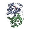
| ||||||||
|---|---|---|---|---|---|---|---|---|---|
| 1 |
| ||||||||
| Unit cell |
|
-Components
| #1: Protein | Mass: 19859.182 Da / Num. of mol.: 2 / Fragment: N-terminal domain / Mutation: V52A Source method: isolated from a genetically manipulated source Source: (gene. exp.) References: UniProt: P31663, pantoate-beta-alanine ligase (AMP-forming) #2: Chemical |   Mass: 147.149 Da / Num. of mol.: 3 / Source method: obtained synthetically / Formula: C6H11O4 Mass: 147.149 Da / Num. of mol.: 3 / Source method: obtained synthetically / Formula: C6H11O4#3: Water | ChemComp-HOH / |  Mass: 18.015 Da / Num. of mol.: 251 / Source method: isolated from a natural source / Formula: H2O Mass: 18.015 Da / Num. of mol.: 251 / Source method: isolated from a natural source / Formula: H2O |
|---|
-Experimental details
-Experiment
| Experiment | Method: X-RAY DIFFRACTION / Number of used crystals: 1 |
|---|
- Sample preparation
| Crystal | Density Matthews: 2.38 Å3/Da / Density % sol: 48.33 % |
|---|---|
| Crystal grow | Temperature: 298 K / Method: vapor diffusion, hanging drop / pH: 5 Details: 10% PEG 4000, 100mM NaCl, 100mM acetate buffer, pH 5.0, VAPOR DIFFUSION, HANGING DROP, temperature 298K |
-Data collection
| Diffraction | Mean temperature: 100 K |
|---|---|
| Diffraction source | Source: ROTATING ANODE / Type: BRUKER AXS MICROSTAR / Wavelength: 1.5418 Å |
| Detector | Type: MAR scanner 345 mm plate / Detector: IMAGE PLATE / Date: Feb 10, 2009 / Details: HELIOS |
| Radiation | Protocol: SINGLE WAVELENGTH / Monochromatic (M) / Laue (L): M / Scattering type: x-ray |
| Radiation wavelength | Wavelength: 1.5418 Å / Relative weight: 1 |
| Reflection | Resolution: 1.67→34.78 Å / Num. obs: 43231 / % possible obs: 96.2 % / Redundancy: 2.5 % / Biso Wilson estimate: 20.3 Å2 / Rmerge(I) obs: 0.051 / Net I/σ(I): 12.8 |
| Reflection shell | Resolution: 1.67→1.76 Å / Redundancy: 2.4 % / Rmerge(I) obs: 0.464 / Mean I/σ(I) obs: 2 / Num. unique all: 5676 / % possible all: 87.5 |
- Processing
| Software |
| ||||||||||||||||||||||||||||||||||||||||||||||||||||||||||||||||||||||||||||||||
|---|---|---|---|---|---|---|---|---|---|---|---|---|---|---|---|---|---|---|---|---|---|---|---|---|---|---|---|---|---|---|---|---|---|---|---|---|---|---|---|---|---|---|---|---|---|---|---|---|---|---|---|---|---|---|---|---|---|---|---|---|---|---|---|---|---|---|---|---|---|---|---|---|---|---|---|---|---|---|---|---|---|
| Refinement | Method to determine structure: MOLECULAR REPLACEMENT / Resolution: 1.67→30.56 Å / Cor.coef. Fo:Fc: 0.949 / Cor.coef. Fo:Fc free: 0.92 / SU B: 4.45 / SU ML: 0.068 / Cross valid method: THROUGHOUT / ESU R: 0.143 / ESU R Free: 0.112 / Stereochemistry target values: MAXIMUM LIKELIHOOD / Details: HYDROGENS HAVE BEEN ADDED IN THE RIDING POSITIONS
| ||||||||||||||||||||||||||||||||||||||||||||||||||||||||||||||||||||||||||||||||
| Solvent computation | Ion probe radii: 0.8 Å / Shrinkage radii: 0.8 Å / VDW probe radii: 1.4 Å / Solvent model: MASK | ||||||||||||||||||||||||||||||||||||||||||||||||||||||||||||||||||||||||||||||||
| Displacement parameters | Biso mean: 11.614 Å2
| ||||||||||||||||||||||||||||||||||||||||||||||||||||||||||||||||||||||||||||||||
| Refinement step | Cycle: LAST / Resolution: 1.67→30.56 Å
| ||||||||||||||||||||||||||||||||||||||||||||||||||||||||||||||||||||||||||||||||
| Refine LS restraints |
| ||||||||||||||||||||||||||||||||||||||||||||||||||||||||||||||||||||||||||||||||
| LS refinement shell | Resolution: 1.669→1.713 Å / Total num. of bins used: 20
| ||||||||||||||||||||||||||||||||||||||||||||||||||||||||||||||||||||||||||||||||
| Refinement TLS params. | Method: refined / Refine-ID: X-RAY DIFFRACTION
| ||||||||||||||||||||||||||||||||||||||||||||||||||||||||||||||||||||||||||||||||
| Refinement TLS group |
|
