 Movie
Movie Controller


+ Open data

- Basic information
| Entry | Database: PDB / ID: 2jd4 | ||||||
|---|---|---|---|---|---|---|---|
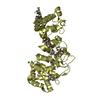
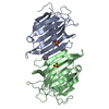
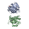
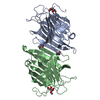
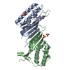
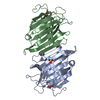
| Title | Mouse laminin alpha1 chain, domains LG4-5 | ||||||
 Components Components | LAMININ SUBUNIT ALPHA-1 | ||||||
 Keywords Keywords | METAL BINDING PROTEIN / LAMININ-111 / BASEMENT MEMBRANE PROTEIN | ||||||
| Function / homology |  Function and homology information Function and homology informationlaminin-121 trimer / laminin trimer / laminin-111 trimer / regulation of basement membrane organization / retinal blood vessel morphogenesis / morphogenesis of an epithelial sheet / glycosphingolipid binding / positive regulation of integrin-mediated signaling pathway / branching involved in salivary gland morphogenesis / tissue development ...laminin-121 trimer / laminin trimer / laminin-111 trimer / regulation of basement membrane organization / retinal blood vessel morphogenesis / morphogenesis of an epithelial sheet / glycosphingolipid binding / positive regulation of integrin-mediated signaling pathway / branching involved in salivary gland morphogenesis / tissue development / positive regulation of skeletal muscle acetylcholine-gated channel clustering / protein complex involved in cell-matrix adhesion / establishment of epithelial cell apical/basal polarity / camera-type eye development / blood vessel morphogenesis / positive regulation of muscle cell differentiation / extracellular matrix structural constituent / epithelial tube branching involved in lung morphogenesis / basement membrane / regulation of embryonic development / positive regulation of Rac protein signal transduction / regulation of cell migration / positive regulation of cell adhesion / axon guidance / animal organ morphogenesis / neuron projection development / cell-cell junction / extracellular matrix / retina development in camera-type eye / positive regulation of phosphatidylinositol 3-kinase/protein kinase B signal transduction / cell surface receptor signaling pathway / cell adhesion / receptor ligand activity / : / extracellular region / membrane Similarity search - Function | ||||||
| Biological species |  | ||||||
| Method |  X-RAY DIFFRACTION / SYNCHROTRON / MOLECULAR REPLACEMENT / Resolution: 1.9 Å X-RAY DIFFRACTION / SYNCHROTRON / MOLECULAR REPLACEMENT / Resolution: 1.9 Å | ||||||
 Authors Authors | Harrison, D. / Hussain, S.A. / Combs, A.C. / Ervasti, J.M. / Yurchenco, P.D. / Hohenester, E. | ||||||
 Citation Citation | Journal: J.Biol.Chem. / Year: 2007 Title: Crystal Structure and Cell Surface Anchorage Sites of Laminin {Alpha}1Lg4-5. Authors: Harrison, D. / Hussain, S.A. / Combs, A.C. / Ervasti, J.M. / Yurchenco, P.D. / Hohenester, E. #1: Journal: Embo J. / Year: 2000Title: Structure of the C-Terminal Laminin G-Like Domain Pair of the Laminin Alpha2 Chain Harbouring Binding Sites for Alpha-Dystroglycan and Heparin Authors: Tisi, D. / Talts, J.F. / Timpl, R. / Hohenester, E. #2: Journal: Annu.Rev.Cell Dev.Biol. / Year: 2004 Title: Laminin Functions in Tissue Morphogenesis Authors: Miner, J.H. / Yurchenco, P.D. | ||||||
| History |
| ||||||
| Remark 700 | SHEET THE SHEET STRUCTURE OF THIS MOLECULE IS BIFURCATED. IN ORDER TO REPRESENT THIS FEATURE IN ... SHEET THE SHEET STRUCTURE OF THIS MOLECULE IS BIFURCATED. IN ORDER TO REPRESENT THIS FEATURE IN THE SHEET RECORDS BELOW, TWO SHEETS ARE DEFINED. |
- Structure visualization
| Structure viewer | Molecule: MolmilJmol/JSmol |
|---|
- Downloads & links
-Download
| PDBx/mmCIF format | 2jd4.cif.gz | 161.7 KB | Display | PDBx/mmCIF format |
|---|---|---|---|---|
| PDB format | pdb2jd4.ent.gz | 127.3 KB | Display | PDB format |
| PDBx/mmJSON format | 2jd4.json.gz | Tree view | PDBx/mmJSON format | |
| Others |  Other downloads Other downloads |
-Validation report
| Arichive directory | https://data.pdbj.org/pub/pdb/validation_reports/jd/2jd4ftp://data.pdbj.org/pub/pdb/validation_reports/jd/2jd4 | HTTPS FTP |
|---|
-Related structure data
| Related structure data |  1dykS S: Starting model for refinement |
|---|---|
| Similar structure data |


-Links
 PDBj
PDBj


- Assembly
| Deposited unit | 
| ||||||||
|---|---|---|---|---|---|---|---|---|---|
| 1 | 
| ||||||||
| 2 | 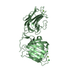
| ||||||||
| Unit cell |
|
-Components
| #1: Protein | Mass: 41901.543 Da / Num. of mol.: 2 / Fragment: DOMAINS LG4-5, RESIDUES 2706-3084 / Mutation: YES Source method: isolated from a genetically manipulated source Source: (gene. exp.)  HOMO SAPIENS (human) / References: UniProt: P19137 HOMO SAPIENS (human) / References: UniProt: P19137#2: Chemical | ChemComp-MG /   Mass: 24.305 Da / Num. of mol.: 4 / Source method: obtained synthetically / Formula: Mg Mass: 24.305 Da / Num. of mol.: 4 / Source method: obtained synthetically / Formula: Mg#3: Chemical |   Mass: 35.453 Da / Num. of mol.: 3 / Source method: obtained synthetically / Formula: Cl Mass: 35.453 Da / Num. of mol.: 3 / Source method: obtained synthetically / Formula: Cl#4: Water | ChemComp-HOH / |  Mass: 18.015 Da / Num. of mol.: 327 / Source method: isolated from a natural source / Formula: H2O Mass: 18.015 Da / Num. of mol.: 327 / Source method: isolated from a natural source / Formula: H2OCompound details | ENGINEERED RESIDUE IN CHAIN A, ASN 2738 TO GLN ENGINEERED RESIDUE IN CHAIN A, ASN 2835 TO LYS ...ENGINEERED | Has protein modification | Y | Sequence details | VECTOR-DERIVED APLA SEQUENCE AT N-TERMINUS, GLYCOSYLATION SITES AND LONE CYSTEINE MUTATED ...VECTOR-DERIVED APLA SEQUENCE AT N-TERMINUS, GLYCOSYLAT | |
|---|
-Experimental details
-Experiment
| Experiment | Method: X-RAY DIFFRACTION / Number of used crystals: 1 |
|---|
- Sample preparation
| Crystal | Density Matthews: 2.3 Å3/Da / Density % sol: 46.02 % |
|---|---|
| Crystal grow | pH: 8.5 / Details: pH 8.50 |
-Data collection
| Diffraction | Mean temperature: 100 K |
|---|---|
| Diffraction source | Source: SYNCHROTRON / Site: SRS  / Beamline: PX9.6 / Wavelength: 0.87 / Beamline: PX9.6 / Wavelength: 0.87 |
| Detector | Type: ADSC CCD / Detector: CCD / Date: May 5, 2005 |
| Radiation | Protocol: SINGLE WAVELENGTH / Monochromatic (M) / Laue (L): M / Scattering type: x-ray |
| Radiation wavelength | Wavelength: 0.87 Å / Relative weight: 1 |
| Reflection | Resolution: 1.9→20 Å / Num. obs: 156499 / % possible obs: 97.3 % / Observed criterion σ(I): 4 / Redundancy: 2.6 % / Rmerge(I) obs: 0.07 / Net I/σ(I): 10.2 |
| Reflection shell | Resolution: 1.9→2 Å / Redundancy: 2.1 % / Rmerge(I) obs: 0.33 / Mean I/σ(I) obs: 2.2 / % possible all: 91.5 |
- Processing
| Software |
| ||||||||||||||||||||||||||||||||||||||||||||||||||||||||||||||||||||||||||||||||
|---|---|---|---|---|---|---|---|---|---|---|---|---|---|---|---|---|---|---|---|---|---|---|---|---|---|---|---|---|---|---|---|---|---|---|---|---|---|---|---|---|---|---|---|---|---|---|---|---|---|---|---|---|---|---|---|---|---|---|---|---|---|---|---|---|---|---|---|---|---|---|---|---|---|---|---|---|---|---|---|---|---|
| Refinement | Method to determine structure: MOLECULAR REPLACEMENT Starting model: PDB ENTRY 1DYK Resolution: 1.9→20 Å / Data cutoff high absF: 10000 / Isotropic thermal model: RESTRAINED / Cross valid method: THROUGHOUT / σ(F): 0 / Stereochemistry target values: MAXIMUM LIKELIHOOD
| ||||||||||||||||||||||||||||||||||||||||||||||||||||||||||||||||||||||||||||||||
| Solvent computation | Solvent model: FLAT MODEL / Bsol: 46.1583 Å2 / ksol: 0.334241 e/Å3 | ||||||||||||||||||||||||||||||||||||||||||||||||||||||||||||||||||||||||||||||||
| Displacement parameters |
| ||||||||||||||||||||||||||||||||||||||||||||||||||||||||||||||||||||||||||||||||
| Refinement step | Cycle: LAST / Resolution: 1.9→20 Å
| ||||||||||||||||||||||||||||||||||||||||||||||||||||||||||||||||||||||||||||||||
| Refine LS restraints |
| ||||||||||||||||||||||||||||||||||||||||||||||||||||||||||||||||||||||||||||||||
| LS refinement shell | Resolution: 1.9→1.91 Å / Total num. of bins used: 50
|
