 Movie
Movie Controller


+ Open data

- Basic information
| Entry | Database: PDB / ID: 2gme | |||||||||
|---|---|---|---|---|---|---|---|---|---|---|
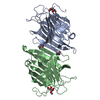
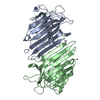
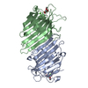
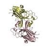
| Title | Metal-free (apo) P. angolensis seed lectin | |||||||||
 Components Components | lectin | |||||||||
 Keywords Keywords | SUGAR BINDING PROTEIN / legume lectin / metal-free lectin / beta sandwich | |||||||||
| Function / homology |  Function and homology information Function and homology information | |||||||||
| Biological species |  Pterocarpus angolensis (plant) Pterocarpus angolensis (plant) | |||||||||
| Method |  X-RAY DIFFRACTION / SYNCHROTRON / MOLECULAR REPLACEMENT / Resolution: 1.75 Å X-RAY DIFFRACTION / SYNCHROTRON / MOLECULAR REPLACEMENT / Resolution: 1.75 Å | |||||||||
 Authors Authors | Garcia-Pino, A. / Buts, L. / Wyns, L. / Loris, R. | |||||||||
 Citation Citation | Journal: J.Mol.Biol. / Year: 2006 Title: Interplay Between Metal Binding and cis/trans Isomerization in Legume Lectins: Structural and Thermodynamic Study of P. angolensis Lectin. Authors: Garcia-Pino, A. / Buts, L. / Wyns, L. / Loris, R. | |||||||||
| History |
|
- Structure visualization
| Structure viewer | Molecule: MolmilJmol/JSmol |
|---|
- Downloads & links
-Download
| PDBx/mmCIF format | 2gme.cif.gz | 107.5 KB | Display | PDBx/mmCIF format |
|---|---|---|---|---|
| PDB format | pdb2gme.ent.gz | 81.7 KB | Display | PDB format |
| PDBx/mmJSON format | 2gme.json.gz | Tree view | PDBx/mmJSON format | |
| Others |  Other downloads Other downloads |
-Validation report
| Arichive directory | https://data.pdbj.org/pub/pdb/validation_reports/gm/2gmeftp://data.pdbj.org/pub/pdb/validation_reports/gm/2gme | HTTPS FTP |
|---|
-Related structure data
| Related structure data | 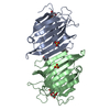 2gmmC 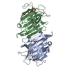 2gmpC  2gn3C 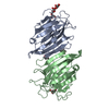 2gn7C 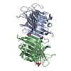 2gnbC 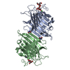 2gndC 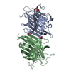 2gnmC 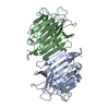 2gntC 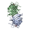 1ukgS S: Starting model for refinement C: citing same article ( |
|---|---|
| Similar structure data |


-Links
 PDBj
PDBj

- Assembly
| Deposited unit | 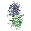
| ||||||||
|---|---|---|---|---|---|---|---|---|---|
| 1 |
| ||||||||
| Unit cell |
| ||||||||
| Details | Subunits A and B together form the lectin dimer |
-Components
| #1: Protein | Mass: 27558.297 Da / Num. of mol.: 2 / Source method: isolated from a natural source / Source: (natural) Pterocarpus angolensis (plant) / Tissue: seed / References: UniProt: Q8GSD2#2: Chemical |   Mass: 96.063 Da / Num. of mol.: 2 / Source method: obtained synthetically / Formula: SO4 Mass: 96.063 Da / Num. of mol.: 2 / Source method: obtained synthetically / Formula: SO4#3: Water | ChemComp-HOH / |  Mass: 18.015 Da / Num. of mol.: 348 / Source method: isolated from a natural source / Formula: H2O Mass: 18.015 Da / Num. of mol.: 348 / Source method: isolated from a natural source / Formula: H2OHas protein modification | Y | |
|---|
-Experimental details
-Experiment
| Experiment | Method: X-RAY DIFFRACTION / Number of used crystals: 1 |
|---|
- Sample preparation
| Crystal | Density Matthews: 2.15 Å3/Da / Density % sol: 42.9 % |
|---|---|
| Crystal grow | Temperature: 293 K / Method: vapor diffusion, hanging drop / pH: 7.2 Details: 10% PEG 6000, 20% MPD, pH 7.2, VAPOR DIFFUSION, HANGING DROP, temperature 293K |
-Data collection
| Diffraction | Mean temperature: 100 K |
|---|---|
| Diffraction source | Source: SYNCHROTRON / Site: EMBL/DESY, HAMBURG  / Beamline: X11 / Wavelength: 0.8123 Å / Beamline: X11 / Wavelength: 0.8123 Å |
| Detector | Type: MAR CCD 165 mm / Detector: CCD |
| Radiation | Monochromator: SAGITALLY FOCUSED Si(111) / Protocol: SINGLE WAVELENGTH / Monochromatic (M) / Laue (L): M / Scattering type: x-ray |
| Radiation wavelength | Wavelength: 0.8123 Å / Relative weight: 1 |
| Reflection | Resolution: 1.75→15 Å / Num. all: 48243 / Num. obs: 48243 / % possible obs: 98 % / Observed criterion σ(F): 0 / Observed criterion σ(I): 0 |
| Reflection shell | Resolution: 1.75→1.81 Å / % possible all: 93.3 |
- Processing
| Software |
| ||||||||||||||||||||
|---|---|---|---|---|---|---|---|---|---|---|---|---|---|---|---|---|---|---|---|---|---|
| Refinement | Method to determine structure: MOLECULAR REPLACEMENT Starting model: pdb entry 1UKG Resolution: 1.75→15 Å / σ(F): 0 / σ(I): 0 / Stereochemistry target values: Engh & Huber
| ||||||||||||||||||||
| Refinement step | Cycle: LAST / Resolution: 1.75→15 Å
| ||||||||||||||||||||
| Refine LS restraints |
|
