 Movie
Movie Controller


+ Open data

- Basic information
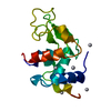
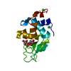
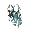
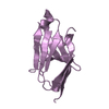
| Entry | Database: PDB / ID: 2axi | ||||||
|---|---|---|---|---|---|---|---|
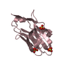
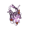
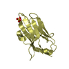
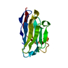
| Title | HDM2 in complex with a beta-hairpin | ||||||
 Components Components |
| ||||||
 Keywords Keywords | LIGASE/LIGASE INHIBITOR / P53 / DRUG DESIGN / PROTEIN-PROTEIN INTERACTIONS / LIGASE / LIGASE-LIGASE INHIBITOR complex | ||||||
| Function / homology |  Function and homology information Function and homology informationcellular response to vitamin B1 / response to formaldehyde / response to water-immersion restraint stress / response to ether / traversing start control point of mitotic cell cycle / atrial septum development / fibroblast activation / regulation of protein catabolic process at postsynapse, modulating synaptic transmission / Trafficking of AMPA receptors / receptor serine/threonine kinase binding ...cellular response to vitamin B1 / response to formaldehyde / response to water-immersion restraint stress / response to ether / traversing start control point of mitotic cell cycle / atrial septum development / fibroblast activation / regulation of protein catabolic process at postsynapse, modulating synaptic transmission / Trafficking of AMPA receptors / receptor serine/threonine kinase binding / negative regulation of intrinsic apoptotic signaling pathway by p53 class mediator / negative regulation of protein processing / response to steroid hormone / SUMO transferase activity / peroxisome proliferator activated receptor binding / positive regulation of vascular associated smooth muscle cell migration / atrioventricular valve morphogenesis / AKT phosphorylates targets in the cytosol / response to iron ion / NEDD8 ligase activity / endocardial cushion morphogenesis / ventricular septum development / cellular response to peptide hormone stimulus / positive regulation of muscle cell differentiation / regulation of postsynaptic neurotransmitter receptor internalization / cardiac septum morphogenesis / SUMOylation of ubiquitinylation proteins / cellular response to alkaloid / blood vessel development / ligase activity / Constitutive Signaling by AKT1 E17K in Cancer / negative regulation of DNA damage response, signal transduction by p53 class mediator / cellular response to antibiotic / SUMOylation of transcription factors / negative regulation of signal transduction by p53 class mediator / regulation of protein catabolic process / cellular response to UV-C / cellular response to estrogen stimulus / protein sumoylation / response to magnesium ion / blood vessel remodeling / ribonucleoprotein complex binding / protein localization to nucleus / protein autoubiquitination / positive regulation of vascular associated smooth muscle cell proliferation / NPAS4 regulates expression of target genes / transcription repressor complex / positive regulation of mitotic cell cycle / regulation of heart rate / : / positive regulation of protein export from nucleus / response to cocaine / ubiquitin binding / DNA damage response, signal transduction by p53 class mediator / establishment of protein localization / sperm end piece / Stabilization of p53 / cellular response to gamma radiation / Regulation of RUNX3 expression and activity / RING-type E3 ubiquitin transferase / Oncogene Induced Senescence / Regulation of TP53 Activity through Methylation / protein destabilization / Degradation of CDH1 / cellular response to growth factor stimulus / response to toxic substance / centriolar satellite / cellular response to hydrogen peroxide / disordered domain specific binding / protein polyubiquitination / p53 binding / ubiquitin-protein transferase activity / endocytic vesicle membrane / Signaling by ALK fusions and activated point mutants / Regulation of TP53 Degradation / ubiquitin protein ligase activity / positive regulation of proteasomal ubiquitin-dependent protein catabolic process / negative regulation of neuron projection development / sperm principal piece / 5S rRNA binding / protein-containing complex assembly / sperm midpiece / Oxidative Stress Induced Senescence / cellular response to hypoxia / Regulation of TP53 Activity through Phosphorylation / amyloid fibril formation / ubiquitin-dependent protein catabolic process / proteasome-mediated ubiquitin-dependent protein catabolic process / regulation of cell cycle / postsynaptic density / Ub-specific processing proteases / protein ubiquitination / response to xenobiotic stimulus / protein domain specific binding / response to antibiotic / negative regulation of DNA-templated transcription / apoptotic process / positive regulation of cell population proliferation / ubiquitin protein ligase binding / positive regulation of gene expression Similarity search - Function | ||||||
| Biological species |  Homo sapiens (human) Homo sapiens (human) | ||||||
| Method |  X-RAY DIFFRACTION / MOLECULAR REPLACEMENT / Resolution: 1.4 Å X-RAY DIFFRACTION / MOLECULAR REPLACEMENT / Resolution: 1.4 Å | ||||||
 Authors Authors | Mittl, P.R.E. / Fasan, R. / Robinson, J. / Gruetter, M.G. | ||||||
 Citation Citation | Journal: Chembiochem / Year: 2006 Title: Structure-Activity Studies in a Family of beta-Hairpin Protein Epitope Mimetic Inhibitors of the p53-HDM2 Protein-Protein Interaction. Authors: Fasan, R. / Dias, R.L. / Moehle, K. / Zerbe, O. / Obrecht, D. / Mittl, P.R. / Robinson, J.A. | ||||||
| History |
|
- Structure visualization
| Structure viewer | Molecule: MolmilJmol/JSmol |
|---|
- Downloads & links
-Download
| PDBx/mmCIF format | 2axi.cif.gz | 41.2 KB | Display | PDBx/mmCIF format |
|---|---|---|---|---|
| PDB format | pdb2axi.ent.gz | 26.9 KB | Display | PDB format |
| PDBx/mmJSON format | 2axi.json.gz | Tree view | PDBx/mmJSON format | |
| Others |  Other downloads Other downloads |
-Validation report
| Arichive directory | https://data.pdbj.org/pub/pdb/validation_reports/ax/2axiftp://data.pdbj.org/pub/pdb/validation_reports/ax/2axi | HTTPS FTP |
|---|
-Related structure data
| Related structure data |  1ycrS S: Starting model for refinement |
|---|---|
| Similar structure data |


-Links
 PDBj
PDBj


- Assembly
| Deposited unit | 
| ||||||||||||
|---|---|---|---|---|---|---|---|---|---|---|---|---|---|
| 1 |
| ||||||||||||
| 2 | 
| ||||||||||||
| 3 | 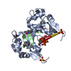
| ||||||||||||
| 4 | 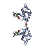
| ||||||||||||
| 5 | 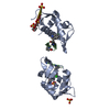
| ||||||||||||
| Unit cell |
| ||||||||||||
| Components on special symmetry positions |
|
-Components
| #1: Protein | Mass: 13364.231 Da / Num. of mol.: 1 / Fragment: HDM2 Source method: isolated from a genetically manipulated source Source: (gene. exp.) Homo sapiens (human) / Gene: MDM2 / Plasmid: pET14b / Production host:  References: UniProt: Q9UMT8, UniProt: Q00987*PLUS, Ligases; Forming carbon-nitrogen bonds; Acid-amino-acid ligases (peptide synthases) | ||||||
|---|---|---|---|---|---|---|---|
| #2: Protein/peptide | 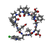  Type: Peptide-like / Class: Inhibitor / Mass: 1399.930 Da / Num. of mol.: 1 / Source method: obtained synthetically / Details: The peptide was synthesized. Type: Peptide-like / Class: Inhibitor / Mass: 1399.930 Da / Num. of mol.: 1 / Source method: obtained synthetically / Details: The peptide was synthesized.References: cyclo(L-alpha-aspartyl-L-tryptophyl-L-alpha-glutamyl-L-phenylalanyl-D-prolyl-L-prolyl-L-phenylalanyl-L-alpha-glutamyl-6 -chloro-L-tryptophyl-L-leucyl) | ||||||
| #3: Chemical |   Mass: 96.063 Da / Num. of mol.: 2 / Source method: obtained synthetically / Formula: SO4 Mass: 96.063 Da / Num. of mol.: 2 / Source method: obtained synthetically / Formula: SO4#4: Chemical | ChemComp-MPO / |   Mass: 209.263 Da / Num. of mol.: 1 / Source method: obtained synthetically / Formula: C7H15NO4S / Comment: pH buffer*YM Mass: 209.263 Da / Num. of mol.: 1 / Source method: obtained synthetically / Formula: C7H15NO4S / Comment: pH buffer*YM#5: Water | ChemComp-HOH / |  Mass: 18.015 Da / Num. of mol.: 96 / Source method: isolated from a natural source / Formula: H2O Mass: 18.015 Da / Num. of mol.: 96 / Source method: isolated from a natural source / Formula: H2OHas protein modification | Y | |
-Experimental details
-Experiment
| Experiment | Method: X-RAY DIFFRACTION / Number of used crystals: 1 |
|---|
- Sample preparation
| Crystal | Density Matthews: 1.82115 Å3/Da / Density % sol: 32.460266 % Description: Data was collected in-house and at the synchrotron |
|---|---|
| Crystal grow | Temperature: 293 K / Method: vapor diffusion, hanging drop / pH: 5.5 Details: MES, ammonium sulfate, pH 5.5, VAPOR DIFFUSION, HANGING DROP, temperature 293K |
-Data collection
| Diffraction | Mean temperature: 100 K |
|---|---|
| Diffraction source | Source: ROTATING ANODE / Type: ENRAF-NONIUS FR571 / Wavelength: 1.54 Å |
| Detector | Type: MARRESEARCH / Detector: IMAGE PLATE / Date: Oct 1, 2003 / Details: Osmic Mirrors |
| Radiation | Protocol: SINGLE WAVELENGTH / Monochromatic (M) / Laue (L): M / Scattering type: x-ray |
| Radiation wavelength | Wavelength: 1.54 Å / Relative weight: 1 |
| Reflection | Resolution: 1.4→50 Å / Num. obs: 21288 / % possible obs: 98.4 % / Observed criterion σ(I): 0 / Redundancy: 4.18 % / Rmerge(I) obs: 0.09 / Net I/σ(I): 15.19 |
| Reflection shell | Resolution: 1.4→1.41 Å / Redundancy: 3.9 % / Rmerge(I) obs: 0.48 / Mean I/σ(I) obs: 2.91 / % possible all: 90.8 |
- Processing
| Software |
| ||||||||||||||||||||||||||||
|---|---|---|---|---|---|---|---|---|---|---|---|---|---|---|---|---|---|---|---|---|---|---|---|---|---|---|---|---|---|
| Refinement | Method to determine structure: MOLECULAR REPLACEMENT Starting model: 1YCR Resolution: 1.4→10 Å / σ(F): 4 / Stereochemistry target values: ENGH & HUBER
| ||||||||||||||||||||||||||||
| Refinement step | Cycle: LAST / Resolution: 1.4→10 Å
|
