 Movie
Movie Controller


+ Open data

- Basic information
| Entry | Database: PDB / ID: 1qc5 | ||||||
|---|---|---|---|---|---|---|---|
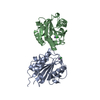
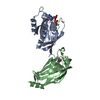
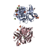
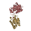
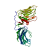
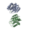
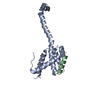
| Title | I Domain from Integrin Alpha1-Beta1 | ||||||
 Components Components | (PROTEIN (ALPHA1 BETA1 INTEGRIN)) x 2 | ||||||
 Keywords Keywords | CELL ADHESION / INTEGRIN | ||||||
| Function / homology |  Function and homology information Function and homology informationintegrin alpha1-beta1 complex / cellular extravasation / collagen binding involved in cell-matrix adhesion / Other semaphorin interactions / CHL1 interactions / phosphatase activator activity / Laminin interactions / basal part of cell / Platelet Adhesion to exposed collagen / integrin complex ...integrin alpha1-beta1 complex / cellular extravasation / collagen binding involved in cell-matrix adhesion / Other semaphorin interactions / CHL1 interactions / phosphatase activator activity / Laminin interactions / basal part of cell / Platelet Adhesion to exposed collagen / integrin complex / negative regulation of epidermal growth factor receptor signaling pathway / Smooth Muscle Contraction / Integrin cell surface interactions / collagen binding / neutrophil chemotaxis / neuron projection morphogenesis / acrosomal vesicle / cell-matrix adhesion / integrin-mediated signaling pathway / cell-cell adhesion / vasodilation / positive regulation of neuron apoptotic process / signaling receptor activity / protein phosphatase binding / perikaryon / positive regulation of MAPK cascade / signaling receptor binding / external side of plasma membrane / negative regulation of cell population proliferation / focal adhesion / cell surface / extracellular exosome / membrane / metal ion binding / plasma membrane Similarity search - Function | ||||||
| Biological species |  Homo sapiens (human) Homo sapiens (human) | ||||||
| Method |  X-RAY DIFFRACTION / MOLECULAR REPLACEMENT / Resolution: 2 Å X-RAY DIFFRACTION / MOLECULAR REPLACEMENT / Resolution: 2 Å | ||||||
 Authors Authors | Deivanayagam, C.C. / Narayana, S.V. | ||||||
 Citation Citation | Journal: J.Biol.Chem. / Year: 1999 Title: Trench-shaped binding sites promote multiple classes of interactions between collagen and the adherence receptors, alpha(1)beta(1) integrin and Staphylococcus aureus cna MSCRAMM. Authors: Rich, R.L. / Deivanayagam, C.C. / Owens, R.T. / Carson, M. / Hook, A. / Moore, D. / Symersky, J. / Yang, V.W. / Narayana, S.V. / Hook, M. | ||||||
| History |
|
- Structure visualization
| Structure viewer | Molecule: MolmilJmol/JSmol |
|---|
- Downloads & links
-Download
| PDBx/mmCIF format | 1qc5.cif.gz | 93.1 KB | Display | PDBx/mmCIF format |
|---|---|---|---|---|
| PDB format | pdb1qc5.ent.gz | 69.3 KB | Display | PDB format |
| PDBx/mmJSON format | 1qc5.json.gz | Tree view | PDBx/mmJSON format | |
| Others |  Other downloads Other downloads |
-Validation report
| Arichive directory | https://data.pdbj.org/pub/pdb/validation_reports/qc/1qc5ftp://data.pdbj.org/pub/pdb/validation_reports/qc/1qc5 | HTTPS FTP |
|---|
-Related structure data
| Related structure data | |
|---|---|
| Similar structure data |


-Links
 PDBj
PDBj


- Assembly
| Deposited unit | 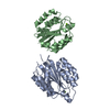
| ||||||||
|---|---|---|---|---|---|---|---|---|---|
| 1 |
| ||||||||
| Unit cell |
| ||||||||
| Noncrystallographic symmetry (NCS) | NCS oper: (Code: given Matrix: (0.99992, -0.00578, -0.01165), Vector: |
-Components
| #1: Protein | Mass: 21501.279 Da / Num. of mol.: 1 / Fragment: I-DOMAIN / Mutation: T137R, Q138S, P139S, T338G Source method: isolated from a genetically manipulated source Source: (gene. exp.) Homo sapiens (human) / Plasmid: PQE-30 / Production host:  | ||
|---|---|---|---|
| #2: Protein | Mass: 21513.334 Da / Num. of mol.: 1 / Fragment: I-DOMAIN / Mutation: T137R, Q138S, P139S, T338G Source method: isolated from a genetically manipulated source Source: (gene. exp.) Homo sapiens (human) / Plasmid: PQE-30 / Production host: | ||
| #3: Chemical |   Mass: 24.305 Da / Num. of mol.: 2 / Source method: obtained synthetically / Formula: Mg Mass: 24.305 Da / Num. of mol.: 2 / Source method: obtained synthetically / Formula: Mg#4: Water | ChemComp-HOH / |  Mass: 18.015 Da / Num. of mol.: 229 / Source method: isolated from a natural source / Formula: H2O Mass: 18.015 Da / Num. of mol.: 229 / Source method: isolated from a natural source / Formula: H2O |
-Experimental details
-Experiment
| Experiment | Method: X-RAY DIFFRACTION / Number of used crystals: 1 |
|---|
- Sample preparation
| Crystal | Density Matthews: 2.16 Å3/Da / Density % sol: 43.17 % | ||||||||||||||||||||||||||||||||||||||||||||||||||||||||||||||||||
|---|---|---|---|---|---|---|---|---|---|---|---|---|---|---|---|---|---|---|---|---|---|---|---|---|---|---|---|---|---|---|---|---|---|---|---|---|---|---|---|---|---|---|---|---|---|---|---|---|---|---|---|---|---|---|---|---|---|---|---|---|---|---|---|---|---|---|---|
| Crystal grow | Temperature: 298 K / Method: vapor diffusion, hanging drop / pH: 7.5 Details: MMEPEG2000, HEPES, SODIUM CHLORIDE, MAGNESIUM CHLORIDE, BETA-MERCAPTOETHANOL, pH 7.50, VAPOR DIFFUSION, HANGING DROP, temperature 298.00K | ||||||||||||||||||||||||||||||||||||||||||||||||||||||||||||||||||
| Crystal | *PLUS Density % sol: 35.3 % | ||||||||||||||||||||||||||||||||||||||||||||||||||||||||||||||||||
| Crystal grow | *PLUS pH: 7 | ||||||||||||||||||||||||||||||||||||||||||||||||||||||||||||||||||
| Components of the solutions | *PLUS
|
-Data collection
| Diffraction | Mean temperature: 100 K |
|---|---|
| Diffraction source | Source: ROTATING ANODE / Type: RIGAKU RUH3R / Wavelength: 1.5418 |
| Detector | Type: RIGAKU RAXIS IV / Detector: IMAGE PLATE / Date: Nov 9, 1998 / Details: MIRRORS |
| Radiation | Monochromator: NI FILTER / Protocol: SINGLE WAVELENGTH / Monochromatic (M) / Laue (L): M / Scattering type: x-ray |
| Radiation wavelength | Wavelength: 1.5418 Å / Relative weight: 1 |
| Reflection | Resolution: 2→100 Å / Num. all: 140776 / Num. obs: 24698 / % possible obs: 99.2 % / Observed criterion σ(F): 1 / Observed criterion σ(I): 1 / Redundancy: 5.7 % / Biso Wilson estimate: 17.1 Å2 / Rmerge(I) obs: 0.061 / Rsym value: 0.061 / Net I/σ(I): 24.1 |
| Reflection shell | Resolution: 1.99→2.07 Å / Redundancy: 4.5 % / Rmerge(I) obs: 0.209 / Mean I/σ(I) obs: 8.1 / Rsym value: 0.209 / % possible all: 95.3 |
| Reflection | *PLUS Highest resolution: 2 Å / Lowest resolution: 100 Å / % possible obs: 99.2 % / Observed criterion σ(F): 1 / Observed criterion σ(I): 1 / Redundancy: 5.7 % / Num. measured all: 140776 / Biso Wilson estimate: 17.1 Å2 |
| Reflection shell | *PLUS Highest resolution: 1.99 Å / Lowest resolution: 2.07 Å / Redundancy: 4.5 % / Mean I/σ(I) obs: 8.1 |
- Processing
| Software |
| ||||||||||||||||||||||||||||||||||||||||||||||||||||||||||||
|---|---|---|---|---|---|---|---|---|---|---|---|---|---|---|---|---|---|---|---|---|---|---|---|---|---|---|---|---|---|---|---|---|---|---|---|---|---|---|---|---|---|---|---|---|---|---|---|---|---|---|---|---|---|---|---|---|---|---|---|---|---|
| Refinement | Method to determine structure: MOLECULAR REPLACEMENT Starting model: THE A DOMAIN OF HUMAN COMPLEMENT FACTOR B Resolution: 2→100 Å / Rfactor Rfree error: 0.005 / Data cutoff high absF: 1166602.56 / Data cutoff low absF: 0 / Isotropic thermal model: RESTRAINED / Cross valid method: THROUGHOUT / σ(F): 2 / σ(I): 4 / Stereochemistry target values: ENGH & HUBER
| ||||||||||||||||||||||||||||||||||||||||||||||||||||||||||||
| Solvent computation | Solvent model: FLAT MODEL / Bsol: 39.29 Å2 / ksol: 0.355 e/Å3 | ||||||||||||||||||||||||||||||||||||||||||||||||||||||||||||
| Displacement parameters | Biso mean: 23.8 Å2
| ||||||||||||||||||||||||||||||||||||||||||||||||||||||||||||
| Refine analyze |
| ||||||||||||||||||||||||||||||||||||||||||||||||||||||||||||
| Refinement step | Cycle: LAST / Resolution: 2→100 Å
| ||||||||||||||||||||||||||||||||||||||||||||||||||||||||||||
| Refine LS restraints |
| ||||||||||||||||||||||||||||||||||||||||||||||||||||||||||||
| LS refinement shell | Resolution: 2→2.13 Å / Rfactor Rfree error: 0.01 / Total num. of bins used: 6
| ||||||||||||||||||||||||||||||||||||||||||||||||||||||||||||
| Xplor file |
| ||||||||||||||||||||||||||||||||||||||||||||||||||||||||||||
| Software | *PLUS Name: CNS / Version: 0.4 / Classification: refinement | ||||||||||||||||||||||||||||||||||||||||||||||||||||||||||||
| Refinement | *PLUS Highest resolution: 2 Å / Lowest resolution: 100 Å / Rfactor obs: 0.206 | ||||||||||||||||||||||||||||||||||||||||||||||||||||||||||||
| Solvent computation | *PLUS | ||||||||||||||||||||||||||||||||||||||||||||||||||||||||||||
| Displacement parameters | *PLUS | ||||||||||||||||||||||||||||||||||||||||||||||||||||||||||||
| LS refinement shell | *PLUS Rfactor Rfree: 0.278 / Rfactor Rwork: 0.232 |
