[English] 日本語
Yorodumi
- PDB-2wrz: Crystal structure of an arabinose binding protein with designed s... -

+
Open data


ID or keywords:

Loading...

-
Basic information

Entry
Database: PDB / ID: 2wrz
TitleCrystal structure of an arabinose binding protein with designed serotonin binding site in open, ligand-free state
ComponentsL-ARABINOSE-BINDING PERIPLASMIC PROTEIN
KeywordsTRANSPORT PROTEIN / SUGAR TRANSPORT / ARABINOSE BINDING PROTEIN / PERIPLASMIC BINDING PROTEIN / ABP / PERIPLASM / RECEPTOR DESIGN
Function / homology
Function and homology information


L-arabinose transmembrane transport / ABC-type monosaccharide transporter activity / monosaccharide binding / ATP-binding cassette (ABC) transporter complex, substrate-binding subunit-containing / outer membrane-bounded periplasmic space / carbohydrate binding / membrane
Similarity search - Function
L-arabinose-binding periplasmic protein / Periplasmic binding protein/LacI sugar binding domain / Periplasmic binding proteins and sugar binding domain of LacI family / Response regulator / Periplasmic binding protein-like I / Rossmann fold / 3-Layer(aba) Sandwich / Alpha Beta
Similarity search - Domain/homology
L-arabinose-binding periplasmic protein
Similarity search - Component
Biological speciesESCHERICHIA COLI (E. coli)
MethodX-RAY DIFFRACTION / SYNCHROTRON / MOLECULAR REPLACEMENT / Resolution: 2.2 Å
AuthorsSchreier, B. / Stumpp, C. / Wiesner, S. / Hocker, B.
CitationJournal: Proc.Natl.Acad.Sci.USA / Year: 2009
Title: The Computational Design of Ligand Binding is not a Solved Problem
Authors: Schreier, B. / Stumpp, C. / Wiesner, S. / Hocker, B.
History
DepositionSep 3, 2009Deposition site: PDBE / Processing site: PDBE
Revision 1.0Oct 13, 2009Provider: repository / Type: Initial release
Revision 1.1May 8, 2011Group: Version format compliance
Revision 1.2Jul 13, 2011Group: Version format compliance
Revision 1.3Dec 20, 2023Group: Data collection / Database references ...Data collection / Database references / Other / Refinement description
Category: chem_comp_atom / chem_comp_bond ...chem_comp_atom / chem_comp_bond / database_2 / pdbx_database_status / pdbx_initial_refinement_model
Item: _database_2.pdbx_DOI / _database_2.pdbx_database_accession / _pdbx_database_status.status_code_sf

-
Structure visualization

Structure viewerMolecule:
MolmilJmol/JSmol

Downloads & links

-
Assembly

Deposited unit
A: L-ARABINOSE-BINDING PERIPLASMIC PROTEIN
B: L-ARABINOSE-BINDING PERIPLASMIC PROTEIN


Theoretical massNumber of molelcules
Total (without water)66,0052
Polymers66,0052
Non-polymers00
Water7,674426
1
A: L-ARABINOSE-BINDING PERIPLASMIC PROTEIN


Theoretical massNumber of molelcules
Total (without water)33,0031
Polymers33,0031
Non-polymers00
Water181
TypeNameSymmetry operationNumber
identity operation1_555x,y,z1
MethodPISA
2
B: L-ARABINOSE-BINDING PERIPLASMIC PROTEIN


Theoretical massNumber of molelcules
Total (without water)33,0031
Polymers33,0031
Non-polymers00
Water181
TypeNameSymmetry operationNumber
identity operation1_555x,y,z1
MethodPISA
Unit cell
Length a, b, c (Å)79.760, 86.300, 116.820
Angle α, β, γ (deg.)90.00, 90.00, 90.00
Int Tables number19
Space group name H-MP212121

-
Components

#1: Protein L-ARABINOSE-BINDING PERIPLASMIC PROTEIN / ABP / ARABINOSE BINDING PROTEIN


Mass: 33002.547 Da / Num. of mol.: 2 / Mutation: YES
Source method: isolated from a genetically manipulated source
Source: (gene. exp.) ESCHERICHIA COLI (E. coli) / Strain: K12 / Plasmid: PET-21 / Production host: ESCHERICHIA COLI (E. coli) / Strain (production host): BL21 / References: UniProt: P02924
#2: Water ChemComp-HOH / water / Water


Mass: 18.015 Da / Num. of mol.: 426 / Source method: isolated from a natural source / Formula: H2O
Compound details
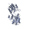
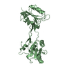
ENGINEERED RESIDUE IN CHAIN A, LYS 33 TO ALA ENGINEERED RESIDUE IN CHAIN A, TRP 39 TO ALA ...ENGINEERED RESIDUE IN CHAIN A, LYS 33 TO ALA ENGINEERED RESIDUE IN CHAIN A, TRP 39 TO ALA ENGINEERED RESIDUE IN CHAIN A, PHE 40 TO SER ENGINEERED RESIDUE IN CHAIN A, GLU 43 TO GLN ENGINEERED RESIDUE IN CHAIN A, CYS 87 TO SER ENGINEERED RESIDUE IN CHAIN A, ASP 113 TO ASN ENGINEERED RESIDUE IN CHAIN A, MET 131 TO GLU ENGINEERED RESIDUE IN CHAIN A, ASN 200 TO GLU ENGINEERED RESIDUE IN CHAIN A, LEU 321 TO CYS ENGINEERED RESIDUE IN CHAIN B, LYS 33 TO ALA ENGINEERED RESIDUE IN CHAIN B, TRP 39 TO ALA ENGINEERED RESIDUE IN CHAIN B, PHE 40 TO SER ENGINEERED RESIDUE IN CHAIN B, GLU 43 TO GLN ENGINEERED RESIDUE IN CHAIN B, CYS 87 TO SER ENGINEERED RESIDUE IN CHAIN B, ASP 113 TO ASN ENGINEERED RESIDUE IN CHAIN B, MET 131 TO GLU ENGINEERED RESIDUE IN CHAIN B, ASN 200 TO GLU ENGINEERED RESIDUE IN CHAIN B, LEU 321 TO CYS

-
Experimental details

-
Experiment

ExperimentMethod: X-RAY DIFFRACTION / Number of used crystals: 1

-
Sample preparation

CrystalDensity Matthews: 2.9 Å3/Da / Density % sol: 58 % / Description: NONE
Crystal growpH: 8
Details: 20% PEG 3350, 0.2M (NH4)CITRATE, 2UMOL/ML SEROTONIN, pH 8.0

-
Data collection

DiffractionMean temperature: 100 K
Diffraction sourceSource: SYNCHROTRON / Site: SLS / Beamline: X10SA / Wavelength: 0.978
DetectorType: MARRESEARCH / Detector: CCD / Date: Jan 27, 2008
RadiationProtocol: SINGLE WAVELENGTH / Monochromatic (M) / Laue (L): M / Scattering type: x-ray
Radiation wavelengthWavelength: 0.978 Å / Relative weight: 1
ReflectionResolution: 2.2→40 Å / Num. obs: 41190 / % possible obs: 99.6 % / Observed criterion σ(I): 2 / Redundancy: 7.27 % / Biso Wilson estimate: 35.593 Å2 / Rmerge(I) obs: 0.17 / Net I/σ(I): 14.8
Reflection shellResolution: 2.2→2.34 Å / Redundancy: 7.08 % / Rmerge(I) obs: 0.87 / Mean I/σ(I) obs: 2.33 / % possible all: 97.8

-
Processing

Software
NameVersionClassification
REFMAC5.4.0066refinement
XDSdata reduction
XDSdata scaling
CCP4Iphasing
RefinementMethod to determine structure: MOLECULAR REPLACEMENT
Starting model: PDB ENTRY 5ABP
Resolution: 2.2→37.96 Å / Cor.coef. Fo:Fc: 0.942 / Cor.coef. Fo:Fc free: 0.904 / SU B: 6.494 / SU ML: 0.164 / Cross valid method: THROUGHOUT / σ(F): 2 / ESU R: 0.237 / ESU R Free: 0.216 / Stereochemistry target values: MAXIMUM LIKELIHOOD / Details: HYDROGENS HAVE BEEN ADDED IN THE RIDING POSITIONS.
RfactorNum. reflection% reflectionSelection details
Rfree0.27159 2093 5 %RANDOM
Rwork0.20994 ---
obs0.21301 39440 99.84 %-
Solvent computationIon probe radii: 0.8 Å / Shrinkage radii: 0.8 Å / VDW probe radii: 1.2 Å / Solvent model: MASK
Displacement parametersBiso mean: 35.35 Å2
Baniso -1Baniso -2Baniso -3
1-0.01 Å20 Å20 Å2
2---0.03 Å20 Å2
3---0.02 Å2
Refinement stepCycle: LAST / Resolution: 2.2→37.96 Å
ProteinNucleic acidLigandSolventTotal
Num. atoms4470 0 0 426 4896
Refine LS restraints
Refine-IDTypeDev idealDev ideal targetNumber
X-RAY DIFFRACTIONr_bond_refined_d0.0120.0224649
X-RAY DIFFRACTIONr_bond_other_d
X-RAY DIFFRACTIONr_angle_refined_deg1.2481.9646317
X-RAY DIFFRACTIONr_angle_other_deg
X-RAY DIFFRACTIONr_dihedral_angle_1_deg5.6375618
X-RAY DIFFRACTIONr_dihedral_angle_2_deg39.71725.692195
X-RAY DIFFRACTIONr_dihedral_angle_3_deg16.16415809
X-RAY DIFFRACTIONr_dihedral_angle_4_deg16.4821518
X-RAY DIFFRACTIONr_chiral_restr0.0830.2709
X-RAY DIFFRACTIONr_gen_planes_refined0.0050.0213524
X-RAY DIFFRACTIONr_gen_planes_other
X-RAY DIFFRACTIONr_nbd_refined
X-RAY DIFFRACTIONr_nbd_other
X-RAY DIFFRACTIONr_nbtor_refined
X-RAY DIFFRACTIONr_nbtor_other
X-RAY DIFFRACTIONr_xyhbond_nbd_refined
X-RAY DIFFRACTIONr_xyhbond_nbd_other
X-RAY DIFFRACTIONr_metal_ion_refined
X-RAY DIFFRACTIONr_metal_ion_other
X-RAY DIFFRACTIONr_symmetry_vdw_refined
X-RAY DIFFRACTIONr_symmetry_vdw_other
X-RAY DIFFRACTIONr_symmetry_hbond_refined
X-RAY DIFFRACTIONr_symmetry_hbond_other
X-RAY DIFFRACTIONr_symmetry_metal_ion_refined
X-RAY DIFFRACTIONr_symmetry_metal_ion_other
X-RAY DIFFRACTIONr_mcbond_it0.7291.52990
X-RAY DIFFRACTIONr_mcbond_other
X-RAY DIFFRACTIONr_mcangle_it1.36424810
X-RAY DIFFRACTIONr_mcangle_other
X-RAY DIFFRACTIONr_scbond_it1.96531659
X-RAY DIFFRACTIONr_scbond_other
X-RAY DIFFRACTIONr_scangle_it3.2694.51493
X-RAY DIFFRACTIONr_scangle_other
X-RAY DIFFRACTIONr_long_range_B_refined
X-RAY DIFFRACTIONr_long_range_B_other
X-RAY DIFFRACTIONr_rigid_bond_restr
X-RAY DIFFRACTIONr_sphericity_free
X-RAY DIFFRACTIONr_sphericity_bonded
LS refinement shellResolution: 2.2→2.257 Å / Total num. of bins used: 20
RfactorNum. reflection% reflection
Rfree0.348 145 -
Rwork0.296 2797 -
obs--98.07 %

+
About Yorodumi

-
News

-
Feb 9, 2022. New format data for meta-information of EMDB entries

New format data for meta-information of EMDB entries

  • Version 3 of the EMDB header file is now the official format.
  • The previous official version 1.9 will be removed from the archive.

Related info.:EMDB header

External links:wwPDB to switch to version 3 of the EMDB data model

-
Aug 12, 2020. Covid-19 info

Covid-19 info

URL: https://pdbj.org/emnavi/covid19.php

New page: Covid-19 featured information page in EM Navigator.

Related info.:Covid-19 info / Mar 5, 2020. Novel coronavirus structure data

+
Mar 5, 2020. Novel coronavirus structure data

Novel coronavirus structure data

Related info.:Yorodumi Speices / Aug 12, 2020. Covid-19 info

External links:COVID-19 featured content - PDBj / Molecule of the Month (242):Coronavirus Proteases

+
Jan 31, 2019. EMDB accession codes are about to change! (news from PDBe EMDB page)

EMDB accession codes are about to change! (news from PDBe EMDB page)

  • The allocation of 4 digits for EMDB accession codes will soon come to an end. Whilst these codes will remain in use, new EMDB accession codes will include an additional digit and will expand incrementally as the available range of codes is exhausted. The current 4-digit format prefixed with “EMD-” (i.e. EMD-XXXX) will advance to a 5-digit format (i.e. EMD-XXXXX), and so on. It is currently estimated that the 4-digit codes will be depleted around Spring 2019, at which point the 5-digit format will come into force.
  • The EM Navigator/Yorodumi systems omit the EMD- prefix.

Related info.:Q: What is EMD? / ID/Accession-code notation in Yorodumi/EM Navigator

External links:EMDB Accession Codes are Changing Soon! / Contact to PDBj

+
Jul 12, 2017. Major update of PDB

Major update of PDB

  • wwPDB released updated PDB data conforming to the new PDBx/mmCIF dictionary.
  • This is a major update changing the version number from 4 to 5, and with Remediation, in which all the entries are updated.
  • In this update, many items about electron microscopy experimental information are reorganized (e.g. em_software).
  • Now, EM Navigator and Yorodumi are based on the updated data.

External links:wwPDB Remediation / Enriched Model Files Conforming to OneDep Data Standards Now Available in the PDB FTP Archive

-
Yorodumi

Thousand views of thousand structures

  • Yorodumi is a browser for structure data from EMDB, PDB, SASBDB, etc.
  • This page is also the successor to EM Navigator detail page, and also detail information page/front-end page for Omokage search.
  • The word "yorodu" (or yorozu) is an old Japanese word meaning "ten thousand". "mi" (miru) is to see.

Related info.:EMDB / PDB / SASBDB / Comparison of 3 databanks / Yorodumi Search / Aug 31, 2016. New EM Navigator & Yorodumi / Yorodumi Papers / Jmol/JSmol / Function and homology information / Changes in new EM Navigator and Yorodumi

Read more