 Movie
Movie Controller

[English] 日本語
 Yorodumi
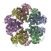
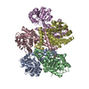
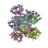
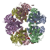
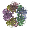
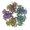
- PDB-6xp3: Structure of human PYCR1 complexed with cyclopentanecarboxylic acid -
+ Open data

- Basic information
| Entry | Database: PDB / ID: 6xp3 | ||||||
|---|---|---|---|---|---|---|---|
| Title | Structure of human PYCR1 complexed with cyclopentanecarboxylic acid | ||||||
 Components Components | Pyrroline-5-carboxylate reductase 1, mitochondrial | ||||||
 Keywords Keywords | OXIDOREDUCTASE / AMINO-ACID BIOSYNTHESIS / PROLINE BIOSYNTHESIS | ||||||
| Function / homology |  Function and homology information Function and homology informationpyrroline-5-carboxylate reductase complex / pyrroline-5-carboxylate reductase / pyrroline-5-carboxylate reductase activity / L-proline biosynthetic process / Glutamate and glutamine metabolism / negative regulation of oxidative stress-induced neuron intrinsic apoptotic signaling pathway / regulation of mitochondrial membrane potential / cellular response to oxidative stress / mitochondrial matrix / mitochondrion / identical protein binding Similarity search - Function | ||||||
| Biological species |  Homo sapiens (human) Homo sapiens (human) | ||||||
| Method |  X-RAY DIFFRACTION / SYNCHROTRON / FOURIER SYNTHESIS / Resolution: 1.93 Å X-RAY DIFFRACTION / SYNCHROTRON / FOURIER SYNTHESIS / Resolution: 1.93 Å | ||||||
 Authors Authors | Tanner, J.J. / Bogner, A.N. | ||||||
| Funding support |  United States, 1items United States, 1items
| ||||||
 Citation Citation | Journal: J.Biol.Chem. / Year: 2020 Title: In crystallo screening for proline analog inhibitors of the proline cycle enzyme PYCR1. Authors: Christensen, E.M. / Bogner, A.N. / Vandekeere, A. / Tam, G.S. / Patel, S.M. / Becker, D.F. / Fendt, S.M. / Tanner, J.J. | ||||||
| History |
|
- Structure visualization
| Structure viewer | Molecule: MolmilJmol/JSmol |
|---|
- Downloads & links
-Download
| PDBx/mmCIF format | 6xp3.cif.gz | 511 KB | Display | PDBx/mmCIF format |
|---|---|---|---|---|
| PDB format | pdb6xp3.ent.gz | 419.5 KB | Display | PDB format |
| PDBx/mmJSON format | 6xp3.json.gz | Tree view | PDBx/mmJSON format | |
| Others |  Other downloads Other downloads |
-Validation report
| Arichive directory | https://data.pdbj.org/pub/pdb/validation_reports/xp/6xp3ftp://data.pdbj.org/pub/pdb/validation_reports/xp/6xp3 | HTTPS FTP |
|---|
-Related structure data
| Related structure data |  6xozC 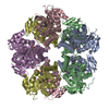 6xp0C 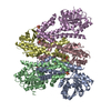 6xp1C  6xp2C  2izzS S: Starting model for refinement C: citing same article ( |
|---|---|
| Similar structure data |


-Links
 PDBj
PDBj


- Assembly
| Deposited unit | 
| ||||||||||||
|---|---|---|---|---|---|---|---|---|---|---|---|---|---|
| 1 | 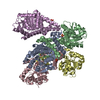
| ||||||||||||
| Unit cell |
| ||||||||||||
| Components on special symmetry positions |
|
-Components
| #1: Protein | Mass: 34043.156 Da / Num. of mol.: 5 Source method: isolated from a genetically manipulated source Source: (gene. exp.) Homo sapiens (human) / Gene: PYCR1 / Production host:  References: UniProt: P32322, pyrroline-5-carboxylate reductase #2: Chemical | ChemComp-SO4 /   Mass: 96.063 Da / Num. of mol.: 4 / Source method: obtained synthetically / Formula: SO4 Mass: 96.063 Da / Num. of mol.: 4 / Source method: obtained synthetically / Formula: SO4#3: Chemical | 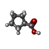  Mass: 114.142 Da / Num. of mol.: 2 / Source method: obtained synthetically / Formula: C6H10O2 / Feature type: SUBJECT OF INVESTIGATION Mass: 114.142 Da / Num. of mol.: 2 / Source method: obtained synthetically / Formula: C6H10O2 / Feature type: SUBJECT OF INVESTIGATION#4: Water | ChemComp-HOH / |  Mass: 18.015 Da / Num. of mol.: 441 / Source method: isolated from a natural source / Formula: H2O Mass: 18.015 Da / Num. of mol.: 441 / Source method: isolated from a natural source / Formula: H2OHas ligand of interest | Y | |
|---|
-Experimental details
-Experiment
| Experiment | Method: X-RAY DIFFRACTION / Number of used crystals: 1 |
|---|
- Sample preparation
| Crystal | Density Matthews: 2.41 Å3/Da / Density % sol: 49.03 % |
|---|---|
| Crystal grow | Temperature: 293 K / Method: vapor diffusion, sitting drop Details: Reservoir contained 200 mM Li2SO4, 18% (w/v) PEG 3350, and 0.1 M HEPES at pH 7.5. Enzyme solution contained 10 mM cyclopentanecarboxylic acid. Crystal was soaked in cryobuffer containing 20% ...Details: Reservoir contained 200 mM Li2SO4, 18% (w/v) PEG 3350, and 0.1 M HEPES at pH 7.5. Enzyme solution contained 10 mM cyclopentanecarboxylic acid. Crystal was soaked in cryobuffer containing 20% PEG 200, and 100 mM cyclopentanecarboxylic acid. |
-Data collection
| Diffraction | Mean temperature: 100 K / Serial crystal experiment: N | ||||||||||||||||||||||||||||||
|---|---|---|---|---|---|---|---|---|---|---|---|---|---|---|---|---|---|---|---|---|---|---|---|---|---|---|---|---|---|---|---|
| Diffraction source | Source: SYNCHROTRON / Site: ALS / Beamline: 4.2.2 / Wavelength: 0.97625 Å | ||||||||||||||||||||||||||||||
| Detector | Type: RDI CMOS_8M / Detector: CMOS / Date: Feb 11, 2020 | ||||||||||||||||||||||||||||||
| Radiation | Protocol: SINGLE WAVELENGTH / Monochromatic (M) / Laue (L): M / Scattering type: x-ray | ||||||||||||||||||||||||||||||
| Radiation wavelength | Wavelength: 0.97625 Å / Relative weight: 1 | ||||||||||||||||||||||||||||||
| Reflection | Resolution: 1.93→47.18 Å / Num. obs: 116598 / % possible obs: 96.7 % / Redundancy: 3.5 % / CC1/2: 0.997 / Rmerge(I) obs: 0.055 / Rpim(I) all: 0.035 / Rrim(I) all: 0.066 / Net I/σ(I): 11.9 / Num. measured all: 408772 / Scaling rejects: 448 | ||||||||||||||||||||||||||||||
| Reflection shell | Diffraction-ID: 1
|
- Processing
| Software |
| ||||||||||||||||||||||||||||||||||||||||||||||||||||||||||||||||||||||||||||||||||||||||||||||||||||||||||||||||||||||||||||||||||||||||||||||||||||||||||||||||||||||||||||||||||||||||||
|---|---|---|---|---|---|---|---|---|---|---|---|---|---|---|---|---|---|---|---|---|---|---|---|---|---|---|---|---|---|---|---|---|---|---|---|---|---|---|---|---|---|---|---|---|---|---|---|---|---|---|---|---|---|---|---|---|---|---|---|---|---|---|---|---|---|---|---|---|---|---|---|---|---|---|---|---|---|---|---|---|---|---|---|---|---|---|---|---|---|---|---|---|---|---|---|---|---|---|---|---|---|---|---|---|---|---|---|---|---|---|---|---|---|---|---|---|---|---|---|---|---|---|---|---|---|---|---|---|---|---|---|---|---|---|---|---|---|---|---|---|---|---|---|---|---|---|---|---|---|---|---|---|---|---|---|---|---|---|---|---|---|---|---|---|---|---|---|---|---|---|---|---|---|---|---|---|---|---|---|---|---|---|---|---|---|---|---|
| Refinement | Method to determine structure: FOURIER SYNTHESIS Starting model: 2IZZ Resolution: 1.93→47.175 Å / SU ML: 0.22 / Cross valid method: THROUGHOUT / σ(F): 1.35 / Phase error: 22.36 / Stereochemistry target values: ML
| ||||||||||||||||||||||||||||||||||||||||||||||||||||||||||||||||||||||||||||||||||||||||||||||||||||||||||||||||||||||||||||||||||||||||||||||||||||||||||||||||||||||||||||||||||||||||||
| Solvent computation | Shrinkage radii: 0.9 Å / VDW probe radii: 1.11 Å / Solvent model: FLAT BULK SOLVENT MODEL | ||||||||||||||||||||||||||||||||||||||||||||||||||||||||||||||||||||||||||||||||||||||||||||||||||||||||||||||||||||||||||||||||||||||||||||||||||||||||||||||||||||||||||||||||||||||||||
| Displacement parameters | Biso max: 108.82 Å2 / Biso mean: 34.9694 Å2 / Biso min: 9.33 Å2 | ||||||||||||||||||||||||||||||||||||||||||||||||||||||||||||||||||||||||||||||||||||||||||||||||||||||||||||||||||||||||||||||||||||||||||||||||||||||||||||||||||||||||||||||||||||||||||
| Refinement step | Cycle: final / Resolution: 1.93→47.175 Å
| ||||||||||||||||||||||||||||||||||||||||||||||||||||||||||||||||||||||||||||||||||||||||||||||||||||||||||||||||||||||||||||||||||||||||||||||||||||||||||||||||||||||||||||||||||||||||||
| LS refinement shell | Refine-ID: X-RAY DIFFRACTION / Rfactor Rfree error: 0
| ||||||||||||||||||||||||||||||||||||||||||||||||||||||||||||||||||||||||||||||||||||||||||||||||||||||||||||||||||||||||||||||||||||||||||||||||||||||||||||||||||||||||||||||||||||||||||
| Refinement TLS params. | Method: refined / Refine-ID: X-RAY DIFFRACTION
| ||||||||||||||||||||||||||||||||||||||||||||||||||||||||||||||||||||||||||||||||||||||||||||||||||||||||||||||||||||||||||||||||||||||||||||||||||||||||||||||||||||||||||||||||||||||||||
| Refinement TLS group |
|
