 Movie
Movie Controller


+ Open data

- Basic information
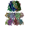
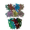
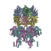
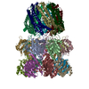
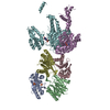
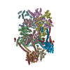
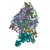
| Entry | Database: PDB / ID: 6xmj | |||||||||
|---|---|---|---|---|---|---|---|---|---|---|
| Title | Human 20S proteasome bound to an engineered 11S (PA26) activator | |||||||||
 Components Components |
| |||||||||
 Keywords Keywords | HYDROLASE / 11S-bound / 20S proteasome | |||||||||
| Function / homology |  Function and homology information Function and homology informationproteasome activator complex / purine ribonucleoside triphosphate binding / Antigen processing: Ub, ATP-independent proteasomal degradation / sperm glycocalyx / Regulation of ornithine decarboxylase (ODC) / Proteasome assembly / perinuclear theca / proteasome core complex / Cross-presentation of soluble exogenous antigens (endosomes) / Somitogenesis ...proteasome activator complex / purine ribonucleoside triphosphate binding / Antigen processing: Ub, ATP-independent proteasomal degradation / sperm glycocalyx / Regulation of ornithine decarboxylase (ODC) / Proteasome assembly / perinuclear theca / proteasome core complex / Cross-presentation of soluble exogenous antigens (endosomes) / Somitogenesis / myofibril / proteasomal ubiquitin-independent protein catabolic process / sperm head-tail coupling apparatus / immune system process / proteasome endopeptidase complex / NF-kappaB binding / proteasome core complex, beta-subunit complex / threonine-type endopeptidase activity / proteasome assembly / proteasome core complex, alpha-subunit complex / regulation of proteasomal protein catabolic process / : / ciliary tip / proteasome complex / sarcomere / Regulation of activated PAK-2p34 by proteasome mediated degradation / Autodegradation of Cdh1 by Cdh1:APC/C / APC/C:Cdc20 mediated degradation of Securin / centriole / Asymmetric localization of PCP proteins / sperm end piece / negative regulation of inflammatory response to antigenic stimulus / Ubiquitin-dependent degradation of Cyclin D / P-body / SCF-beta-TrCP mediated degradation of Emi1 / NIK-->noncanonical NF-kB signaling / AUF1 (hnRNP D0) binds and destabilizes mRNA / lipopolysaccharide binding / TNFR2 non-canonical NF-kB pathway / Assembly of the pre-replicative complex / Vpu mediated degradation of CD4 / Cdc20:Phospho-APC/C mediated degradation of Cyclin A / Dectin-1 mediated noncanonical NF-kB signaling / Degradation of DVL / Degradation of AXIN / Degradation of CRY and PER proteins / Hh mutants are degraded by ERAD / Activation of NF-kappaB in B cells / G2/M Checkpoints / Degradation of GLI1 by the proteasome / Hedgehog ligand biogenesis / Autodegradation of the E3 ubiquitin ligase COP1 / Regulation of RUNX3 expression and activity / Defective CFTR causes cystic fibrosis / Negative regulation of NOTCH4 signaling / GSK3B and BTRC:CUL1-mediated-degradation of NFE2L2 / APC/C:Cdh1 mediated degradation of Cdc20 and other APC/C:Cdh1 targeted proteins in late mitosis/early G1 / Hedgehog 'on' state / Vif-mediated degradation of APOBEC3G / FBXL7 down-regulates AURKA during mitotic entry and in early mitosis / Degradation of GLI2 by the proteasome / GLI3 is processed to GLI3R by the proteasome / Ubiquitin-Mediated Degradation of Phosphorylated Cdc25A / MAPK6/MAPK4 signaling / Degradation of CDH1 / Degradation of beta-catenin by the destruction complex / Oxygen-dependent proline hydroxylation of Hypoxia-inducible Factor Alpha / CDK-mediated phosphorylation and removal of Cdc6 / ABC-family protein mediated transport / CLEC7A (Dectin-1) signaling / SCF(Skp2)-mediated degradation of p27/p21 / FCERI mediated NF-kB activation / response to virus / nuclear matrix / Regulation of expression of SLITs and ROBOs / Regulation of PTEN stability and activity / Interleukin-1 signaling / Orc1 removal from chromatin / Regulation of RUNX2 expression and activity / Regulation of RAS by GAPs / The role of GTSE1 in G2/M progression after G2 checkpoint / Separation of Sister Chromatids / UCH proteinases / KEAP1-NFE2L2 pathway / peptidase activity / Downstream TCR signaling / Antigen processing: Ubiquitination & Proteasome degradation / sperm principal piece / RUNX1 regulates transcription of genes involved in differentiation of HSCs / ER-Phagosome pathway / Neddylation / regulation of inflammatory response / response to oxidative stress / sperm midpiece / secretory granule lumen / endopeptidase activity / ficolin-1-rich granule lumen / proteasome-mediated ubiquitin-dependent protein catabolic process / positive regulation of canonical NF-kappaB signal transduction / Ub-specific processing proteases Similarity search - Function | |||||||||
| Biological species |  Homo sapiens (human) Homo sapiens (human) | |||||||||
| Method | ELECTRON MICROSCOPY / single particle reconstruction / cryo EM / Resolution: 3 Å | |||||||||
 Authors Authors | de la Pena, A.H. / Opoku-Nsiah, K.A. / Williams, S.K. / Chopra, N. / Sali, A. / Gestwicki, J.E. / Lander, G.C. | |||||||||
| Funding support |  United States, 2items United States, 2items
| |||||||||
 Citation Citation | Journal: Nat Commun / Year: 2022 Title: The YΦ motif defines the structure-activity relationships of human 20S proteasome activators. Authors: Kwadwo A Opoku-Nsiah / Andres H de la Pena / Sarah K Williams / Nikita Chopra / Andrej Sali / Gabriel C Lander / Jason E Gestwicki / Abstract: The 20S proteasome (20S) facilitates turnover of most eukaryotic proteins. Substrate entry into the 20S first requires opening of gating loops through binding of HbYX motifs that are present at the C- ...The 20S proteasome (20S) facilitates turnover of most eukaryotic proteins. Substrate entry into the 20S first requires opening of gating loops through binding of HbYX motifs that are present at the C-termini of certain proteasome activators (PAs). The HbYX motif has been predominantly characterized in the archaeal 20S, whereas little is known about the sequence preferences of the human 20S (h20S). Here, we synthesize and screen ~120 HbYX-like peptides, revealing unexpected differences from the archaeal system and defining the h20S recognition sequence as the Y-F/Y (YФ) motif. To gain further insight, we create a functional chimera of the optimized sequence, NLSYYT, fused to the model activator, PA26. A cryo-EM structure of PA26-h20S is used to identify key interactions, including non-canonical contacts and gate-opening mechanisms. Finally, we demonstrate that the YФ sequence preferences are tuned by valency, allowing multivalent PAs to sample greater sequence space. These results expand the model for termini-mediated gating and provide a template for the design of h20S activators. | |||||||||
| History |
|
- Structure visualization
| Movie |
Movie viewer |
|---|---|
| Structure viewer | Molecule: MolmilJmol/JSmol |
- Downloads & links
-Download
| PDBx/mmCIF format | 6xmj.cif.gz | 768.2 KB | Display | PDBx/mmCIF format |
|---|---|---|---|---|
| PDB format | pdb6xmj.ent.gz | 626.4 KB | Display | PDB format |
| PDBx/mmJSON format | 6xmj.json.gz | Tree view | PDBx/mmJSON format | |
| Others |  Other downloads Other downloads |
-Validation report
| Arichive directory | https://data.pdbj.org/pub/pdb/validation_reports/xm/6xmjftp://data.pdbj.org/pub/pdb/validation_reports/xm/6xmj | HTTPS FTP |
|---|
-Related structure data
| Related structure data |  22259MC M: map data used to model this data C: citing same article ( |
|---|---|
| Similar structure data |

-Links
 PDBj
PDBj


- Assembly
| Deposited unit | 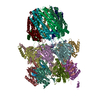
|
|---|---|
| 1 |
|
-Components
-Proteasome subunit alpha type- ... , 7 types, 7 molecules ABCDEFG
| #1: Protein | Mass: 27186.174 Da / Num. of mol.: 1 Source method: isolated from a genetically manipulated source Source: (gene. exp.) Homo sapiens (human) / Gene: PSMA6, PROS27 / Cell (production host): erythrocytes / Production host: Homo sapiens (human)References: UniProt: P60900, proteasome endopeptidase complex |
|---|---|
| #2: Protein | Mass: 25796.338 Da / Num. of mol.: 1 Source method: isolated from a genetically manipulated source Source: (gene. exp.) Homo sapiens (human) / Gene: PSMA2, HC3, PSC3 / Cell (production host): erythrocytes / Production host: Homo sapiens (human)References: UniProt: P25787, proteasome endopeptidase complex |
| #3: Protein | Mass: 28118.189 Da / Num. of mol.: 1 Source method: isolated from a genetically manipulated source Source: (gene. exp.) Homo sapiens (human) / Gene: PSMA4, HC9, PSC9 / Cell (production host): erythrocytes / Production host: Homo sapiens (human)References: UniProt: P25789, proteasome endopeptidase complex |
| #4: Protein | Mass: 27382.178 Da / Num. of mol.: 1 Source method: isolated from a genetically manipulated source Source: (gene. exp.) Homo sapiens (human) / Gene: PSMA7, HSPC / Cell (production host): erythrocytes / Production host: Homo sapiens (human)References: UniProt: O14818, proteasome endopeptidase complex |
| #5: Protein | Mass: 25569.957 Da / Num. of mol.: 1 Source method: isolated from a genetically manipulated source Source: (gene. exp.) Homo sapiens (human) / Gene: PSMA5 / Cell (production host): erythrocytes / Production host: Homo sapiens (human)References: UniProt: P28066, proteasome endopeptidase complex |
| #6: Protein | Mass: 26728.428 Da / Num. of mol.: 1 Source method: isolated from a genetically manipulated source Source: (gene. exp.) Homo sapiens (human) / Gene: PSMA1, HC2, NU, PROS30, PSC2 / Cell (production host): erythrocytes / Production host: Homo sapiens (human)References: UniProt: P25786, proteasome endopeptidase complex |
| #7: Protein | Mass: 27287.100 Da / Num. of mol.: 1 Source method: isolated from a genetically manipulated source Source: (gene. exp.) Homo sapiens (human) / Gene: PSMA3, HC8, PSC8 / Cell (production host): erythrocytes / Production host: Homo sapiens (human)References: UniProt: P25788, proteasome endopeptidase complex |
-Proteasome subunit beta type- ... , 7 types, 7 molecules HIJKLMN
| #8: Protein | Mass: 21656.527 Da / Num. of mol.: 1 Source method: isolated from a genetically manipulated source Source: (gene. exp.) Homo sapiens (human) / Gene: PSMB6, LMPY, Y / Cell (production host): erythrocytes / Production host: Homo sapiens (human)References: UniProt: P28072, proteasome endopeptidase complex |
|---|---|
| #9: Protein | Mass: 23745.256 Da / Num. of mol.: 1 Source method: isolated from a genetically manipulated source Source: (gene. exp.) Homo sapiens (human) / Gene: PSMB7, Z / Cell (production host): erythrocytes / Production host: Homo sapiens (human)References: UniProt: Q99436, proteasome endopeptidase complex |
| #10: Protein | Mass: 22841.701 Da / Num. of mol.: 1 Source method: isolated from a genetically manipulated source Source: (gene. exp.) Homo sapiens (human) / Gene: PSMB3 / Cell (production host): erythrocytes / Production host: Homo sapiens (human)References: UniProt: P49720, proteasome endopeptidase complex |
| #11: Protein | Mass: 22720.146 Da / Num. of mol.: 1 Source method: isolated from a genetically manipulated source Source: (gene. exp.) Homo sapiens (human) / Gene: PSMB2 / Cell (production host): erythrocytes / Production host: Homo sapiens (human)References: UniProt: P49721, proteasome endopeptidase complex |
| #12: Protein | Mass: 22199.072 Da / Num. of mol.: 1 Source method: isolated from a genetically manipulated source Source: (gene. exp.) Homo sapiens (human) / Gene: PSMB5, LMPX, MB1, X / Cell (production host): erythrocytes / Production host: Homo sapiens (human)References: UniProt: P28074, proteasome endopeptidase complex |
| #13: Protein | Mass: 23578.986 Da / Num. of mol.: 1 Source method: isolated from a genetically manipulated source Source: (gene. exp.) Homo sapiens (human) / Gene: PSMB1, PSC5 / Cell (production host): erythrocytes / Production host: Homo sapiens (human)References: UniProt: P20618, proteasome endopeptidase complex |
| #14: Protein | Mass: 24138.453 Da / Num. of mol.: 1 Source method: isolated from a genetically manipulated source Source: (gene. exp.) Homo sapiens (human) / Gene: PSMB4, PROS26 / Cell (production host): erythrocytes / Production host: Homo sapiens (human)References: UniProt: P28070, proteasome endopeptidase complex |
-Protein , 1 types, 7 molecules OPQRSTU
| #15: Protein | Mass: 24959.373 Da / Num. of mol.: 7 Source method: isolated from a genetically manipulated source Source: (gene. exp.)  |
|---|
-Experimental details
-Experiment
| Experiment | Method: ELECTRON MICROSCOPY |
|---|---|
| EM experiment | Aggregation state: PARTICLE / 3D reconstruction method: single particle reconstruction |
- Sample preparation
| Component |
| ||||||||||||||||||||||||||||||
|---|---|---|---|---|---|---|---|---|---|---|---|---|---|---|---|---|---|---|---|---|---|---|---|---|---|---|---|---|---|---|---|
| Source (natural) |
| ||||||||||||||||||||||||||||||
| Source (recombinant) |
| ||||||||||||||||||||||||||||||
| Buffer solution | pH: 7.6 | ||||||||||||||||||||||||||||||
| Buffer component |
| ||||||||||||||||||||||||||||||
| Specimen | Conc.: 20 mg/ml / Embedding applied: NO / Shadowing applied: NO / Staining applied: NO / Vitrification applied: YES | ||||||||||||||||||||||||||||||
| Specimen support | Grid material: COPPER / Grid type: Quantifoil R2/2 | ||||||||||||||||||||||||||||||
| Vitrification | Instrument: HOMEMADE PLUNGER / Cryogen name: ETHANE / Humidity: 90 % / Chamber temperature: 277 K Details: Specimens were manually blotted with Whatman #1 filter paper. |
- Electron microscopy imaging
| Experimental equipment |  Model: Titan Krios / Image courtesy: FEI Company |
|---|---|
| Microscopy | Model: FEI TITAN KRIOS / Details: Images were acquired in nanoprobe mode. |
| Electron gun | Electron source:  FIELD EMISSION GUN / Accelerating voltage: 300 kV / Illumination mode: FLOOD BEAM FIELD EMISSION GUN / Accelerating voltage: 300 kV / Illumination mode: FLOOD BEAM |
| Electron lens | Mode: BRIGHT FIELD / Nominal magnification: 29000 X / Nominal defocus max: -2500 nm / Nominal defocus min: -1000 nm / Calibrated defocus min: -1500 nm / Calibrated defocus max: -3000 nm / Cs: 2.7 mm / C2 aperture diameter: 70 µm / Alignment procedure: COMA FREE |
| Specimen holder | Cryogen: NITROGEN / Specimen holder model: FEI TITAN KRIOS AUTOGRID HOLDER |
| Image recording | Average exposure time: 6.25 sec. / Electron dose: 50 e/Å2 / Detector mode: COUNTING / Film or detector model: GATAN K2 SUMMIT (4k x 4k) / Num. of grids imaged: 1 / Num. of real images: 11656 |
| Image scans | Sampling size: 5 µm / Width: 3710 / Height: 3838 / Movie frames/image: 25 / Used frames/image: 1-25 |
- Processing
| EM software |
| ||||||||||||||||||||||||||||||||||||||||||||||||||||
|---|---|---|---|---|---|---|---|---|---|---|---|---|---|---|---|---|---|---|---|---|---|---|---|---|---|---|---|---|---|---|---|---|---|---|---|---|---|---|---|---|---|---|---|---|---|---|---|---|---|---|---|---|---|
| Image processing | Details: Camera was operated in counting mode. | ||||||||||||||||||||||||||||||||||||||||||||||||||||
| CTF correction | Details: CTF correction was performed by Relion during reconstruction. Type: PHASE FLIPPING AND AMPLITUDE CORRECTION | ||||||||||||||||||||||||||||||||||||||||||||||||||||
| Particle selection | Num. of particles selected: 579361 Details: Particles were selected using the Relion template-based particle picker. | ||||||||||||||||||||||||||||||||||||||||||||||||||||
| Symmetry | Point symmetry: C1 (asymmetric) | ||||||||||||||||||||||||||||||||||||||||||||||||||||
| 3D reconstruction | Resolution: 3 Å / Resolution method: FSC 0.143 CUT-OFF / Num. of particles: 234960 / Symmetry type: POINT | ||||||||||||||||||||||||||||||||||||||||||||||||||||
| Atomic model building | Protocol: RIGID BODY FIT / Space: REAL |
