 Movie
Movie Controller

[English] 日本語
 Yorodumi
- PDB-3jrm: Crystal structure of archaeal 20S proteasome in complex with muta... -
+ Open data

- Basic information
| Entry | Database: PDB / ID: 3jrm | ||||||
|---|---|---|---|---|---|---|---|
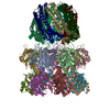
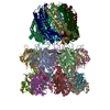
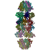
| Title | Crystal structure of archaeal 20S proteasome in complex with mutated P26 activator | ||||||
 Components Components |
| ||||||
 Keywords Keywords | HYDROLASE/HYDROLASE ACTIVATOR / 20S proteasome / PA26 / Cytoplasm / Hydrolase / Protease / Proteasome / Threonine protease / HYDROLASE-HYDROLASE ACTIVATOR COMPLEX | ||||||
| Function / homology |  Function and homology information Function and homology informationproteasome activator complex / proteasome endopeptidase complex / proteasome core complex, beta-subunit complex / threonine-type endopeptidase activity / proteasome core complex, alpha-subunit complex / regulation of proteasomal protein catabolic process / proteasomal protein catabolic process / endopeptidase activity / ubiquitin-dependent protein catabolic process / proteolysis / cytoplasm Similarity search - Function | ||||||
| Biological species |   Thermoplasma acidophilum (acidophilic) Thermoplasma acidophilum (acidophilic) | ||||||
| Method |  X-RAY DIFFRACTION / SYNCHROTRON / MOLECULAR REPLACEMENT / Resolution: 2.9 Å X-RAY DIFFRACTION / SYNCHROTRON / MOLECULAR REPLACEMENT / Resolution: 2.9 Å | ||||||
 Authors Authors | Stadtmueller, B.M. / Whitby, F.G. / Hill, C.P. | ||||||
 Citation Citation | Journal: J.Biol.Chem. / Year: 2010 Title: Structural models for interactions between the 20S proteasome and its PAN/19S activators. Authors: Stadtmueller, B.M. / Ferrell, K. / Whitby, F.G. / Heroux, A. / Robinson, H. / Myszka, D.G. / Hill, C.P. | ||||||
| History |
|
- Structure visualization
| Structure viewer | Molecule: MolmilJmol/JSmol |
|---|
- Downloads & links
-Download
| PDBx/mmCIF format | 3jrm.cif.gz | 818.6 KB | Display | PDBx/mmCIF format |
|---|---|---|---|---|
| PDB format | pdb3jrm.ent.gz | 688 KB | Display | PDB format |
| PDBx/mmJSON format | 3jrm.json.gz | Tree view | PDBx/mmJSON format | |
| Others |  Other downloads Other downloads |
-Validation report
| Arichive directory | https://data.pdbj.org/pub/pdb/validation_reports/jr/3jrmftp://data.pdbj.org/pub/pdb/validation_reports/jr/3jrm | HTTPS FTP |
|---|
-Related structure data
| Related structure data |  3jseC  3jtlC 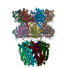 1yarS C: citing same article ( S: Starting model for refinement |
|---|---|
| Similar structure data |


-Links
 PDBj
PDBj


- Assembly
| Deposited unit | 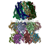
| ||||||||||||||||||||||||||||||||||||||||||||||||||||||||||||||||||||||||||||||||||||||||||||||||||||||||||||||||||||||||||||||||||||||||||||||||||||||||||||||||||||||||||||||||||||||||||||||||||||||||||||
|---|---|---|---|---|---|---|---|---|---|---|---|---|---|---|---|---|---|---|---|---|---|---|---|---|---|---|---|---|---|---|---|---|---|---|---|---|---|---|---|---|---|---|---|---|---|---|---|---|---|---|---|---|---|---|---|---|---|---|---|---|---|---|---|---|---|---|---|---|---|---|---|---|---|---|---|---|---|---|---|---|---|---|---|---|---|---|---|---|---|---|---|---|---|---|---|---|---|---|---|---|---|---|---|---|---|---|---|---|---|---|---|---|---|---|---|---|---|---|---|---|---|---|---|---|---|---|---|---|---|---|---|---|---|---|---|---|---|---|---|---|---|---|---|---|---|---|---|---|---|---|---|---|---|---|---|---|---|---|---|---|---|---|---|---|---|---|---|---|---|---|---|---|---|---|---|---|---|---|---|---|---|---|---|---|---|---|---|---|---|---|---|---|---|---|---|---|---|---|---|---|---|---|---|---|---|
| 1 | 
| ||||||||||||||||||||||||||||||||||||||||||||||||||||||||||||||||||||||||||||||||||||||||||||||||||||||||||||||||||||||||||||||||||||||||||||||||||||||||||||||||||||||||||||||||||||||||||||||||||||||||||||
| Unit cell |
| ||||||||||||||||||||||||||||||||||||||||||||||||||||||||||||||||||||||||||||||||||||||||||||||||||||||||||||||||||||||||||||||||||||||||||||||||||||||||||||||||||||||||||||||||||||||||||||||||||||||||||||
| Noncrystallographic symmetry (NCS) | NCS domain:
NCS domain segments: Component-ID: 1 / Refine code: 1
|