 Movie
Movie Controller

[English] 日本語
 Yorodumi
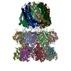
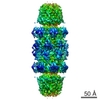
- PDB-1yar: Structure of Archeabacterial 20S proteasome mutant D9S- PA26 complex -
+ Open data

- Basic information
| Entry | Database: PDB / ID: 1yar | ||||||
|---|---|---|---|---|---|---|---|
| Title | Structure of Archeabacterial 20S proteasome mutant D9S- PA26 complex | ||||||
 Components Components |
| ||||||
 Keywords Keywords | HYDROLASE/HYDROLASE ACTIVATOR / proteasome 20S / PA26 proteasome activator 11S / HYDROLASE-HYDROLASE ACTIVATOR COMPLEX | ||||||
| Function / homology |  Function and homology information Function and homology informationproteasome activator complex / proteasome endopeptidase complex / proteasome core complex, beta-subunit complex / threonine-type endopeptidase activity / proteasome core complex, alpha-subunit complex / regulation of proteasomal protein catabolic process / proteasomal protein catabolic process / endopeptidase activity / ubiquitin-dependent protein catabolic process / proteolysis / cytoplasm Similarity search - Function | ||||||
| Biological species |   Thermoplasma acidophilum (acidophilic) Thermoplasma acidophilum (acidophilic) | ||||||
| Method |  X-RAY DIFFRACTION / SYNCHROTRON / MOLECULAR REPLACEMENT / Resolution: 1.9 Å X-RAY DIFFRACTION / SYNCHROTRON / MOLECULAR REPLACEMENT / Resolution: 1.9 Å | ||||||
 Authors Authors | Forster, A. / Masters, E.I. / Whitby, F.G. / Robinson, H. / Hill, C.P. | ||||||
 Citation Citation | Journal: Mol.Cell / Year: 2005 Title: The 1.9 A structure of a proteasome-11S activator complex and implications for proteasome-PAN/PA700 interactions. Authors: Forster, A. / Masters, E.I. / Whitby, F.G. / Robinson, H. / Hill, C.P. | ||||||
| History |
| ||||||
| Remark 999 | SEQUENCE For the Proteasome beta subunit (chains H,I,J,K,L,M,N) the first 8 residues are cleaved ...SEQUENCE For the Proteasome beta subunit (chains H,I,J,K,L,M,N) the first 8 residues are cleaved off autocatalytically during assembly of the complex. |
- Structure visualization
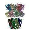
| Structure viewer | Molecule: MolmilJmol/JSmol |
|---|
- Downloads & links
-Download
| PDBx/mmCIF format | 1yar.cif.gz | 945.4 KB | Display | PDBx/mmCIF format |
|---|---|---|---|---|
| PDB format | pdb1yar.ent.gz | 784.5 KB | Display | PDB format |
| PDBx/mmJSON format | 1yar.json.gz | Tree view | PDBx/mmJSON format | |
| Others |  Other downloads Other downloads |
-Validation report
| Arichive directory | https://data.pdbj.org/pub/pdb/validation_reports/ya/1yarftp://data.pdbj.org/pub/pdb/validation_reports/ya/1yar | HTTPS FTP |
|---|
-Related structure data
| Related structure data | 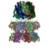 1ya7C  1yauC 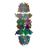 1z7qC 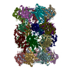 1pmaS C: citing same article ( S: Starting model for refinement |
|---|---|
| Similar structure data |


-Links
 PDBj
PDBj


- Assembly
| Deposited unit | 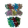
| ||||||||
|---|---|---|---|---|---|---|---|---|---|
| 1 | 
| ||||||||
| Unit cell |
| ||||||||
| Details | T. acidophilum is a 28-subunit barrel-shaped complex. It contains 2 stacked homoheptameric rings of beta subunits flanked by 2 homoheptameric rings of alpha subunits. One half of the 20S complex is contained in the asymmetric unit (one ring of beta and one ring of alpha subunits. / PA26 is a homoheptameric ring that binds to the end of the 20S proteasome through interactions with the 20S alpha subunits. One PA26 complex is contained in the asymmetric unit. |
-Components
-Protein , 3 types, 21 molecules ABCDEFGHIJKLMNOPQRSTU
| #1: Protein | Mass: 25801.439 Da / Num. of mol.: 7 / Mutation: D9S Source method: isolated from a genetically manipulated source Source: (gene. exp.) Thermoplasma acidophilum (acidophilic) / Gene: psmA / Plasmid: pRSET5a / Species (production host): Escherichia coli / Production host:  References: UniProt: P25156, proteasome endopeptidase complex #2: Protein | Mass: 23998.691 Da / Num. of mol.: 7 Source method: isolated from a genetically manipulated source Source: (gene. exp.) Thermoplasma acidophilum (acidophilic) / Gene: psmB / Plasmid: pRSET5a / Species (production host): Escherichia coli / Production host: References: UniProt: P28061, proteasome endopeptidase complex #3: Protein | Mass: 26087.643 Da / Num. of mol.: 7 Source method: isolated from a genetically manipulated source Source: (gene. exp.) |
|---|
-Non-polymers , 3 types, 3498 molecules 


| #4: Chemical | ChemComp-SO4 /  Mass: 96.063 Da / Num. of mol.: 21 / Source method: obtained synthetically / Formula: SO4 Mass: 96.063 Da / Num. of mol.: 21 / Source method: obtained synthetically / Formula: SO4#5: Chemical | ChemComp-GOL /  Mass: 92.094 Da / Num. of mol.: 7 / Source method: obtained synthetically / Formula: C3H8O3 Mass: 92.094 Da / Num. of mol.: 7 / Source method: obtained synthetically / Formula: C3H8O3#6: Water | ChemComp-HOH / | Mass: 18.015 Da / Num. of mol.: 3470 / Source method: isolated from a natural source / Formula: H2O |
|---|
-Experimental details
-Experiment
| Experiment | Method: X-RAY DIFFRACTION / Number of used crystals: 1 |
|---|
- Sample preparation
| Crystal | Density Matthews: 3.4 Å3/Da / Density % sol: 63 % |
|---|---|
| Crystal grow | Temperature: 273 K / Method: vapor diffusion, hanging drop / pH: 4.2 Details: 0.1M Na-citrate/phosphate buffer, 0.2M Lithium Sulfate, 15% PEG-1000, pH 4.2, VAPOR DIFFUSION, HANGING DROP, temperature 273K |
-Data collection
| Diffraction | Mean temperature: 100 K |
|---|---|
| Diffraction source | Source: SYNCHROTRON / Site: NSLS  / Beamline: X25 / Wavelength: 1.283 Å / Beamline: X25 / Wavelength: 1.283 Å |
| Detector | Type: ADSC QUANTUM 4 / Detector: CCD / Date: Feb 22, 2004 |
| Radiation | Protocol: SINGLE WAVELENGTH / Monochromatic (M) / Laue (L): M / Scattering type: x-ray |
| Radiation wavelength | Wavelength: 1.283 Å / Relative weight: 1 |
| Reflection | Resolution: 1.9→20 Å / Num. obs: 442007 / % possible obs: 97.4 % / Observed criterion σ(F): 0 / Observed criterion σ(I): 0 / Redundancy: 5 % / Rmerge(I) obs: 0.063 / Rsym value: 0.063 / Net I/σ(I): 10 |
| Reflection shell | Resolution: 1.9→1.97 Å / Rmerge(I) obs: 0.497 / Mean I/σ(I) obs: 2.2 / % possible all: 95.8 |
- Processing
| Software |
| ||||||||||||||||||||||||||||||||||||||||||||||||||||||||||||||||||||||||||||||||||||||||||
|---|---|---|---|---|---|---|---|---|---|---|---|---|---|---|---|---|---|---|---|---|---|---|---|---|---|---|---|---|---|---|---|---|---|---|---|---|---|---|---|---|---|---|---|---|---|---|---|---|---|---|---|---|---|---|---|---|---|---|---|---|---|---|---|---|---|---|---|---|---|---|---|---|---|---|---|---|---|---|---|---|---|---|---|---|---|---|---|---|---|---|---|
| Refinement | Method to determine structure: MOLECULAR REPLACEMENT Starting model: PDB entry 1PMA Resolution: 1.9→47.3 Å / Cor.coef. Fo:Fc: 0.961 / Cor.coef. Fo:Fc free: 0.945 / SU B: 2.843 / SU ML: 0.085 / Cross valid method: THROUGHOUT / σ(F): 0 / σ(I): 0 / ESU R: 0.131 / ESU R Free: 0.124 / Stereochemistry target values: MAXIMUM LIKELIHOOD / Details: HYDROGENS HAVE BEEN ADDED IN THE RIDING POSITIONS
| ||||||||||||||||||||||||||||||||||||||||||||||||||||||||||||||||||||||||||||||||||||||||||
| Solvent computation | Ion probe radii: 0.8 Å / Shrinkage radii: 0.8 Å / VDW probe radii: 1.2 Å / Solvent model: MASK | ||||||||||||||||||||||||||||||||||||||||||||||||||||||||||||||||||||||||||||||||||||||||||
| Displacement parameters | Biso mean: 31.532 Å2
| ||||||||||||||||||||||||||||||||||||||||||||||||||||||||||||||||||||||||||||||||||||||||||
| Refinement step | Cycle: LAST / Resolution: 1.9→47.3 Å
| ||||||||||||||||||||||||||||||||||||||||||||||||||||||||||||||||||||||||||||||||||||||||||
| Refine LS restraints |
| ||||||||||||||||||||||||||||||||||||||||||||||||||||||||||||||||||||||||||||||||||||||||||
| LS refinement shell | Resolution: 1.899→1.949 Å / Total num. of bins used: 20
|
