 Movie
Movie Controller

[English] 日本語
 Yorodumi
- PDB-1z7q: Crystal structure of the 20s proteasome from yeast in complex wit... -
+ Open data

- Basic information
| Entry | Database: PDB / ID: 1z7q | ||||||
|---|---|---|---|---|---|---|---|
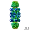
| Title | Crystal structure of the 20s proteasome from yeast in complex with the proteasome activator PA26 from Trypanosome brucei at 3.2 angstroms resolution | ||||||
 Components Components |
| ||||||
 Keywords Keywords | HYDROLASE/HYDROLASE ACTIVATOR / 20S / proteasome / PA26 / activator / multi-catalytic protease / HYDROLASE-HYDROLASE ACTIVATOR COMPLEX | ||||||
| Function / homology |  Function and homology information Function and homology informationproteasome activator complex / ER-Phagosome pathway / Antigen processing: Ub, ATP-independent proteasomal degradation / proteasome core complex assembly / Proteasome assembly / Cross-presentation of soluble exogenous antigens (endosomes) / TNFR2 non-canonical NF-kB pathway / nuclear outer membrane-endoplasmic reticulum membrane network / Regulation of PTEN stability and activity / CDK-mediated phosphorylation and removal of Cdc6 ...proteasome activator complex / ER-Phagosome pathway / Antigen processing: Ub, ATP-independent proteasomal degradation / proteasome core complex assembly / Proteasome assembly / Cross-presentation of soluble exogenous antigens (endosomes) / TNFR2 non-canonical NF-kB pathway / nuclear outer membrane-endoplasmic reticulum membrane network / Regulation of PTEN stability and activity / CDK-mediated phosphorylation and removal of Cdc6 / FBXL7 down-regulates AURKA during mitotic entry and in early mitosis / KEAP1-NFE2L2 pathway / Neddylation / Ubiquitin-Mediated Degradation of Phosphorylated Cdc25A / Orc1 removal from chromatin / MAPK6/MAPK4 signaling / Antigen processing: Ubiquitination & Proteasome degradation / proteasome storage granule / proteasomal ubiquitin-independent protein catabolic process / Ub-specific processing proteases / proteasome endopeptidase complex / proteasome core complex, beta-subunit complex / endopeptidase activator activity / threonine-type endopeptidase activity / proteasome core complex, alpha-subunit complex / proteasome assembly / Neutrophil degranulation / regulation of proteasomal protein catabolic process / proteasome complex / peroxisome / endopeptidase activity / proteasome-mediated ubiquitin-dependent protein catabolic process / mRNA binding / endoplasmic reticulum membrane / mitochondrion / proteolysis / nucleus / cytosol Similarity search - Function | ||||||
| Biological species |   | ||||||
| Method |  X-RAY DIFFRACTION / SYNCHROTRON / MOLECULAR REPLACEMENT / Resolution: 3.22 Å X-RAY DIFFRACTION / SYNCHROTRON / MOLECULAR REPLACEMENT / Resolution: 3.22 Å | ||||||
 Authors Authors | Forster, A. / Whitby, F.G. / Hill, C.P. | ||||||
 Citation Citation | Journal: Mol.Cell / Year: 2005 Title: The 1.9 A structure of a proteasome-11S activator complex and implications for proteasome-PAN/PA700 interactions. Authors: Forster, A. / Masters, E.I. / Whitby, F.G. / Robinson, H. / Hill, C.P. | ||||||
| History |
| ||||||
| Remark 999 | SEQUENCE Residue (i GLY 1172) and Residue (i GLY 1173) are not linked. Distance of C-N bond is 1.91. ...SEQUENCE Residue (i GLY 1172) and Residue (i GLY 1173) are not linked. Distance of C-N bond is 1.91. Residue (p GLY 1172) and Residue (p GLY 1173) are not linked. Distance of C-N bond is 1.91. |
- Structure visualization
| Structure viewer | Molecule: MolmilJmol/JSmol |
|---|
- Downloads & links
-Download
| PDBx/mmCIF format | 1z7q.cif.gz | 1.8 MB | Display | PDBx/mmCIF format |
|---|---|---|---|---|
| PDB format | pdb1z7q.ent.gz | 1.4 MB | Display | PDB format |
| PDBx/mmJSON format | 1z7q.json.gz | Tree view | PDBx/mmJSON format | |
| Others |  Other downloads Other downloads |
-Validation report
| Arichive directory | https://data.pdbj.org/pub/pdb/validation_reports/z7/1z7qftp://data.pdbj.org/pub/pdb/validation_reports/z7/1z7q | HTTPS FTP |
|---|
-Related structure data
| Related structure data |  1ya7C  1yarC  1yauC  1rypS S: Starting model for refinement C: citing same article ( |
|---|---|
| Similar structure data |


-Links
 PDBj
PDBj


- Assembly
| Deposited unit | 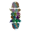
| ||||||||
|---|---|---|---|---|---|---|---|---|---|
| 1 |
| ||||||||
| Unit cell |
| ||||||||
| Details | 1 complete, 28-subunit 20S proteasome is found in the asymmetric unit / 2 complete, 7-subunit PA26 compllexes are found in the asymmetric unit |
-Components
-Proteasome component ... , 13 types, 26 molecules AOBPCQDRESFTGUHVIWJXKYLZNb
| #1: Protein | Mass: 28033.830 Da / Num. of mol.: 2 Source method: isolated from a genetically manipulated source Source: (gene. exp.) Production host: References: UniProt: P21243, proteasome endopeptidase complex #2: Protein | Mass: 27191.828 Da / Num. of mol.: 2 Source method: isolated from a genetically manipulated source Source: (gene. exp.) Production host: References: UniProt: P23639, proteasome endopeptidase complex #3: Protein | Mass: 28748.230 Da / Num. of mol.: 2 Source method: isolated from a genetically manipulated source Source: (gene. exp.) Production host: References: UniProt: P23638, proteasome endopeptidase complex #4: Protein | Mass: 28478.111 Da / Num. of mol.: 2 Source method: isolated from a genetically manipulated source Source: (gene. exp.) Production host: References: UniProt: P40303, proteasome endopeptidase complex #5: Protein | Mass: 28649.086 Da / Num. of mol.: 2 Source method: isolated from a genetically manipulated source Source: (gene. exp.) Production host: References: UniProt: P32379, proteasome endopeptidase complex #6: Protein | Mass: 25634.000 Da / Num. of mol.: 2 Source method: isolated from a genetically manipulated source Source: (gene. exp.) Production host: References: UniProt: P40302, proteasome endopeptidase complex #7: Protein | Mass: 31575.068 Da / Num. of mol.: 2 Source method: isolated from a genetically manipulated source Source: (gene. exp.) Production host: References: UniProt: P21242, proteasome endopeptidase complex #8: Protein | Mass: 21517.186 Da / Num. of mol.: 2 Source method: isolated from a genetically manipulated source Source: (gene. exp.) Production host: References: UniProt: P38624, proteasome endopeptidase complex #9: Protein | Mass: 23987.254 Da / Num. of mol.: 2 Source method: isolated from a genetically manipulated source Source: (gene. exp.) Production host: References: UniProt: P25043, proteasome endopeptidase complex #10: Protein | Mass: 22627.842 Da / Num. of mol.: 2 Source method: isolated from a genetically manipulated source Source: (gene. exp.) Production host: References: UniProt: P25451, proteasome endopeptidase complex #11: Protein | Mass: 22545.676 Da / Num. of mol.: 2 Source method: isolated from a genetically manipulated source Source: (gene. exp.) Production host: References: UniProt: P22141, proteasome endopeptidase complex #12: Protein | Mass: 23353.262 Da / Num. of mol.: 2 Source method: isolated from a genetically manipulated source Source: (gene. exp.) Production host: References: UniProt: P30656, proteasome endopeptidase complex #14: Protein | Mass: 25945.496 Da / Num. of mol.: 2 Source method: isolated from a genetically manipulated source Source: (gene. exp.) Production host: References: UniProt: P30657, proteasome endopeptidase complex |
|---|
-Protein , 2 types, 16 molecules Macdefghijklmnop
| #13: Protein | Mass: 24883.928 Da / Num. of mol.: 2 Source method: isolated from a genetically manipulated source Source: (gene. exp.) Production host: References: UniProt: P23724, proteasome endopeptidase complex #15: Protein | Mass: 25228.734 Da / Num. of mol.: 14 Source method: isolated from a genetically manipulated source Source: (gene. exp.)  |
|---|
-Experimental details
-Experiment
| Experiment | Method: X-RAY DIFFRACTION / Number of used crystals: 1 |
|---|
- Sample preparation
| Crystal | Density Matthews: 3.08 Å3/Da / Density % sol: 60.04 % |
|---|---|
| Crystal grow | Temperature: 298 K / Method: vapor diffusion, sitting drop / pH: 7.5 Details: 0.1 M NaHEPES, 40% MPD, 0.2 M NACL, pH 7.50, VAPOR DIFFUSION, SITTING DROP, temperature 298K |
-Data collection
| Diffraction | Mean temperature: 100 K |
|---|---|
| Diffraction source | Source: SYNCHROTRON / Site: SSRL  / Beamline: BL9-1 / Wavelength: 0.98 Å / Beamline: BL9-1 / Wavelength: 0.98 Å |
| Detector | Type: MARRESEARCH / Detector: IMAGE PLATE / Date: Feb 15, 2000 |
| Radiation | Monochromator: SSRL beamline 9-1 / Protocol: SINGLE WAVELENGTH / Monochromatic (M) / Laue (L): M / Scattering type: x-ray |
| Radiation wavelength | Wavelength: 0.98 Å / Relative weight: 1 |
| Reflection | Resolution: 3.2→50 Å / Num. all: 189615 / Num. obs: 189615 / % possible obs: 89.1 % / Observed criterion σ(F): 0 / Observed criterion σ(I): 0 / Redundancy: 2.7 % / Rmerge(I) obs: 0.112 / Rsym value: 0.112 / Net I/σ(I): 7 |
| Reflection shell | Resolution: 3.2→3.26 Å / Redundancy: 2.5 % / Rmerge(I) obs: 0.331 / Mean I/σ(I) obs: 1.9 / Rsym value: 0.331 / % possible all: 0.0629 |
- Processing
| Software |
| |||||||||||||||||||||||||
|---|---|---|---|---|---|---|---|---|---|---|---|---|---|---|---|---|---|---|---|---|---|---|---|---|---|---|
| Refinement | Method to determine structure: MOLECULAR REPLACEMENT Starting model: one-half of the yeast 20S proteasome (Huber, et al., high-resolution structure), pdb entry 1RYP Resolution: 3.22→39.8 Å / σ(F): 0 / σ(I): 0 / Stereochemistry target values: Engh & Huber
| |||||||||||||||||||||||||
| Refinement step | Cycle: LAST / Resolution: 3.22→39.8 Å
|
