 Movie
Movie Controller


+ Open data

- Basic information
| Entry | Database: PDB / ID: 5ji3 | |||||||||
|---|---|---|---|---|---|---|---|---|---|---|
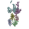
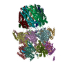
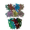
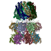
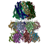
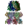
| Title | HslUV complex | |||||||||
 Components Components |
| |||||||||
 Keywords Keywords | HYDROLASE / AAA+ ATPase / peptidase / HSLVU / PEPTIDASE-ATPASE COMPLEX / HYDROLASE COMPLEX | |||||||||
| Function / homology |  Function and homology information Function and homology informationprotein denaturation / HslU-HslV peptidase / HslUV protease complex / proteasome-activating activity / proteasome core complex / protein unfolding / threonine-type endopeptidase activity / : / peptidase activity / cellular response to heat ...protein denaturation / HslU-HslV peptidase / HslUV protease complex / proteasome-activating activity / proteasome core complex / protein unfolding / threonine-type endopeptidase activity / : / peptidase activity / cellular response to heat / response to heat / protein domain specific binding / magnesium ion binding / ATP hydrolysis activity / proteolysis / ATP binding / membrane / metal ion binding / identical protein binding / cytoplasm / cytosol Similarity search - Function | |||||||||
| Biological species |  | |||||||||
| Method |  X-RAY DIFFRACTION / SYNCHROTRON / MOLECULAR REPLACEMENT / Resolution: 3 Å X-RAY DIFFRACTION / SYNCHROTRON / MOLECULAR REPLACEMENT / Resolution: 3 Å | |||||||||
 Authors Authors | Grant, R.A. / Sauer, R.T. / Schmitz, K.R. / Baytshtok, V. | |||||||||
| Funding support |  United States, 2items United States, 2items
| |||||||||
 Citation Citation | Journal: Structure / Year: 2016 Title: A Structurally Dynamic Region of the HslU Intermediate Domain Controls Protein Degradation and ATP Hydrolysis. Authors: Baytshtok, V. / Fei, X. / Grant, R.A. / Baker, T.A. / Sauer, R.T. #1: Journal: Structure / Year: 2001Title: Crystal structures of the HslVU peptidase-ATPase complex reveal an ATP-dependent proteolysis mechanism Authors: Wang, J. / Song, J.J. / Franklin, M.C. / Kamtekar, S. / Im, Y.J. / Rho, S.H. / Seong, I.S. / Lee, C.S. / Chung, C.H. / Eom, S.H. | |||||||||
| History |
|
- Structure visualization
| Structure viewer | Molecule: MolmilJmol/JSmol |
|---|
- Downloads & links
-Download
| PDBx/mmCIF format | 5ji3.cif.gz | 521.6 KB | Display | PDBx/mmCIF format |
|---|---|---|---|---|
| PDB format | pdb5ji3.ent.gz | 434.3 KB | Display | PDB format |
| PDBx/mmJSON format | 5ji3.json.gz | Tree view | PDBx/mmJSON format | |
| Others |  Other downloads Other downloads |
-Validation report
| Arichive directory | https://data.pdbj.org/pub/pdb/validation_reports/ji/5ji3ftp://data.pdbj.org/pub/pdb/validation_reports/ji/5ji3 | HTTPS FTP |
|---|
-Related structure data
| Related structure data | 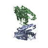 5ji2C 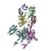 1g4aS S: Starting model for refinement C: citing same article ( |
|---|---|
| Similar structure data |
-Links
 PDBj
PDBj


- Assembly
| Deposited unit | 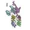
| ||||||||||||||||||||||||||||||
|---|---|---|---|---|---|---|---|---|---|---|---|---|---|---|---|---|---|---|---|---|---|---|---|---|---|---|---|---|---|---|---|
| 1 | 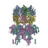
| ||||||||||||||||||||||||||||||
| Unit cell |
| ||||||||||||||||||||||||||||||
| Symmetry | Point symmetry: (Schoenflies symbol: C3 (3 fold cyclic)) | ||||||||||||||||||||||||||||||
| Noncrystallographic symmetry (NCS) | NCS domain:
NCS domain segments:
|
-Components
| #1: Protein | Mass: 19117.836 Da / Num. of mol.: 4 Source method: isolated from a genetically manipulated source Source: (gene. exp.) References: UniProt: B7LA29, UniProt: P0A7B8*PLUS, HslU-HslV peptidase #2: Protein | Mass: 49659.703 Da / Num. of mol.: 2 Source method: isolated from a genetically manipulated source Source: (gene. exp.) #3: Chemical |   Mass: 411.202 Da / Num. of mol.: 2 / Source method: obtained synthetically / Formula: C10H15N5O9P2 Mass: 411.202 Da / Num. of mol.: 2 / Source method: obtained synthetically / Formula: C10H15N5O9P2 |
|---|
-Experimental details
-Experiment
| Experiment | Method: X-RAY DIFFRACTION / Number of used crystals: 1 |
|---|
- Sample preparation
| Crystal | Density Matthews: 4.28 Å3/Da / Density % sol: 71.24 % |
|---|---|
| Crystal grow | Temperature: 300 K / Method: vapor diffusion, hanging drop / Details: NA |
-Data collection
| Diffraction | Mean temperature: 100 K |
|---|---|
| Diffraction source | Source: SYNCHROTRON / Site: ALS / Beamline: 5.0.2 / Wavelength: 0.97 Å |
| Detector | Type: ADSC QUANTUM 315 / Detector: CCD / Date: Feb 22, 2000 |
| Radiation | Protocol: SINGLE WAVELENGTH / Monochromatic (M) / Laue (L): M / Scattering type: x-ray |
| Radiation wavelength | Wavelength: 0.97 Å / Relative weight: 1 |
| Reflection | % possible obs: 85.7 % / Redundancy: 2 % / Net I/σ(I): 2 |
- Processing
| Software |
| |||||||||||||||||||||||||||||||||||||||||||||||||||||||||||||||||||||||||||||||||||||||||||||||||||||||||||||||||||||||||||||||||||||||||||||||||||
|---|---|---|---|---|---|---|---|---|---|---|---|---|---|---|---|---|---|---|---|---|---|---|---|---|---|---|---|---|---|---|---|---|---|---|---|---|---|---|---|---|---|---|---|---|---|---|---|---|---|---|---|---|---|---|---|---|---|---|---|---|---|---|---|---|---|---|---|---|---|---|---|---|---|---|---|---|---|---|---|---|---|---|---|---|---|---|---|---|---|---|---|---|---|---|---|---|---|---|---|---|---|---|---|---|---|---|---|---|---|---|---|---|---|---|---|---|---|---|---|---|---|---|---|---|---|---|---|---|---|---|---|---|---|---|---|---|---|---|---|---|---|---|---|---|---|---|---|---|
| Refinement | Method to determine structure: MOLECULAR REPLACEMENT Starting model: 1G4A Resolution: 3→84.997 Å / Cross valid method: FREE R-VALUE / σ(F): 0 / Phase error: 39.89 Details: data were downloaded as mmCIF file from PDB 1G4A and converted to MTZ format for re-refinement of the model
| |||||||||||||||||||||||||||||||||||||||||||||||||||||||||||||||||||||||||||||||||||||||||||||||||||||||||||||||||||||||||||||||||||||||||||||||||||
| Solvent computation | Shrinkage radii: 0.9 Å / VDW probe radii: 1.11 Å | |||||||||||||||||||||||||||||||||||||||||||||||||||||||||||||||||||||||||||||||||||||||||||||||||||||||||||||||||||||||||||||||||||||||||||||||||||
| Refinement step | Cycle: LAST / Resolution: 3→84.997 Å
| |||||||||||||||||||||||||||||||||||||||||||||||||||||||||||||||||||||||||||||||||||||||||||||||||||||||||||||||||||||||||||||||||||||||||||||||||||
| Refine LS restraints |
| |||||||||||||||||||||||||||||||||||||||||||||||||||||||||||||||||||||||||||||||||||||||||||||||||||||||||||||||||||||||||||||||||||||||||||||||||||
| Refine LS restraints NCS |
| |||||||||||||||||||||||||||||||||||||||||||||||||||||||||||||||||||||||||||||||||||||||||||||||||||||||||||||||||||||||||||||||||||||||||||||||||||
| LS refinement shell |
|
