 Movie
Movie Controller

[English] 日本語
 Yorodumi
- PDB-6x0o: Single-Particle Cryo-EM Structure of Arabinosyltransferase EmbB f... -
+ Open data

- Basic information
| Entry | Database: PDB / ID: 6x0o | |||||||||||||||||||||||||||||||||||||||
|---|---|---|---|---|---|---|---|---|---|---|---|---|---|---|---|---|---|---|---|---|---|---|---|---|---|---|---|---|---|---|---|---|---|---|---|---|---|---|---|---|
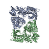
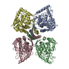
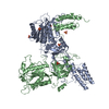
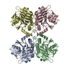
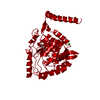
| Title | Single-Particle Cryo-EM Structure of Arabinosyltransferase EmbB from Mycobacterium smegmatis | |||||||||||||||||||||||||||||||||||||||
 Components Components | Integral membrane indolylacetylinositol arabinosyltransferase EmbB | |||||||||||||||||||||||||||||||||||||||
 Keywords Keywords | MEMBRANE PROTEIN / Glycosyltransferase / nanodisc | |||||||||||||||||||||||||||||||||||||||
| Function / homology |  Function and homology information Function and homology informationindolylacetylinositol arabinosyltransferase / indolylacetylinositol arabinosyltransferase activity / arabinosyltransferase activity / Actinobacterium-type cell wall biogenesis / Transferases; Glycosyltransferases; Pentosyltransferases / cell wall organization / plasma membrane Similarity search - Function | |||||||||||||||||||||||||||||||||||||||
| Biological species |  Mycolicibacterium smegmatis (bacteria) Mycolicibacterium smegmatis (bacteria) | |||||||||||||||||||||||||||||||||||||||
| Method | ELECTRON MICROSCOPY / single particle reconstruction / cryo EM / Resolution: 3.3 Å | |||||||||||||||||||||||||||||||||||||||
 Authors Authors | Tan, Y.Z. / Rodrigues, J. / Keener, J.E. / Zheng, R.B. / Brunton, R. / Kloss, B. / Giacometti, S.I. / Rosario, A.L. / Zhang, L. / Niederweis, M. ...Tan, Y.Z. / Rodrigues, J. / Keener, J.E. / Zheng, R.B. / Brunton, R. / Kloss, B. / Giacometti, S.I. / Rosario, A.L. / Zhang, L. / Niederweis, M. / Clarke, O.B. / Lowary, T.L. / Marty, M.T. / Archer, M. / Potter, C.S. / Carragher, B. / Mancia, F. | |||||||||||||||||||||||||||||||||||||||
| Funding support |  United States, United States,  Portugal, European Union, Portugal, European Union,  Canada, 12items Canada, 12items
| |||||||||||||||||||||||||||||||||||||||
 Citation Citation | Journal: Nat Commun / Year: 2020 Title: Cryo-EM structure of arabinosyltransferase EmbB from Mycobacterium smegmatis. Authors: Yong Zi Tan / José Rodrigues / James E Keener / Ruixiang Blake Zheng / Richard Brunton / Brian Kloss / Sabrina I Giacometti / Ana L Rosário / Lei Zhang / Michael Niederweis / Oliver B ...Authors: Yong Zi Tan / José Rodrigues / James E Keener / Ruixiang Blake Zheng / Richard Brunton / Brian Kloss / Sabrina I Giacometti / Ana L Rosário / Lei Zhang / Michael Niederweis / Oliver B Clarke / Todd L Lowary / Michael T Marty / Margarida Archer / Clinton S Potter / Bridget Carragher / Filippo Mancia /  Abstract: Arabinosyltransferase B (EmbB) belongs to a family of membrane-bound glycosyltransferases that build the lipidated polysaccharides of the mycobacterial cell envelope, and are targets of anti- ...Arabinosyltransferase B (EmbB) belongs to a family of membrane-bound glycosyltransferases that build the lipidated polysaccharides of the mycobacterial cell envelope, and are targets of anti-tuberculosis drug ethambutol. We present the 3.3 Å resolution single-particle cryo-electron microscopy structure of Mycobacterium smegmatis EmbB, providing insights on substrate binding and reaction mechanism. Mutations that confer ethambutol resistance map mostly around the putative active site, suggesting this to be the location of drug binding. | |||||||||||||||||||||||||||||||||||||||
| History |
|
- Structure visualization
| Movie |
Movie viewer |
|---|---|
| Structure viewer | Molecule: MolmilJmol/JSmol |
- Downloads & links
-Download
| PDBx/mmCIF format | 6x0o.cif.gz | 345.1 KB | Display | PDBx/mmCIF format |
|---|---|---|---|---|
| PDB format | pdb6x0o.ent.gz | 280.5 KB | Display | PDB format |
| PDBx/mmJSON format | 6x0o.json.gz | Tree view | PDBx/mmJSON format | |
| Others |  Other downloads Other downloads |
-Validation report
| Arichive directory | https://data.pdbj.org/pub/pdb/validation_reports/x0/6x0oftp://data.pdbj.org/pub/pdb/validation_reports/x0/6x0o | HTTPS FTP |
|---|
-Related structure data
| Related structure data |  21983MC M: map data used to model this data C: citing same article ( |
|---|---|
| Similar structure data | |
| EM raw data | EMPIAR-10420 (Title: Single-Particle Cryo-EM of Arabinosyltransferase EmbB from Mycobacterium smegmatis, Collected using both Falcon III and K2 Detectors Data size: 4.5 TB Data #1: Unaligned and compressed multiframe movies from Falcon III Detector [micrographs - multiframe] Data #2: Unaligned and compressed multiframe movies from K2 Summit Detector [micrographs - multiframe] Data #3: Aligned and dose-weighted micrographs from Falcon III Detector [micrographs - single frame] Data #4: Aligned and dose-weighted micrographs from K2 Summit Detector [micrographs - single frame] Data #5: Final Particle Stack with Refined Euler Angles and Shifts [picked particles - single frame - processed]) |

-Links
 PDBj
PDBj


- Assembly
| Deposited unit | 
|
|---|---|
| 1 |
|
-Components
| #1: Protein | Mass: 118835.375 Da / Num. of mol.: 1 Source method: isolated from a genetically manipulated source Source: (gene. exp.) Mycolicibacterium smegmatis (strain ATCC 700084 / mc(2)155) (bacteria)Strain: ATCC 700084 / mc(2)155 / Gene: embB, MSMEI_6221 / Production host: References: UniProt: I7GAQ2, UniProt: A0R614*PLUS, indolylacetylinositol arabinosyltransferase | ||||
|---|---|---|---|---|---|
| #2: Chemical |   Mass: 40.078 Da / Num. of mol.: 2 / Source method: obtained synthetically / Formula: Ca / Feature type: SUBJECT OF INVESTIGATION Mass: 40.078 Da / Num. of mol.: 2 / Source method: obtained synthetically / Formula: Ca / Feature type: SUBJECT OF INVESTIGATION#3: Chemical |   Mass: 722.970 Da / Num. of mol.: 2 / Source method: obtained synthetically / Formula: C38H75O10P / Feature type: SUBJECT OF INVESTIGATION / Comment: phospholipid*YM Mass: 722.970 Da / Num. of mol.: 2 / Source method: obtained synthetically / Formula: C38H75O10P / Feature type: SUBJECT OF INVESTIGATION / Comment: phospholipid*YMHas ligand of interest | Y | |
-Experimental details
-Experiment
| Experiment | Method: ELECTRON MICROSCOPY |
|---|---|
| EM experiment | Aggregation state: PARTICLE / 3D reconstruction method: single particle reconstruction |
- Sample preparation
| Component | Name: Integral membrane indolylacetylinositol arabinosyltransferase EmbB Type: ORGANELLE OR CELLULAR COMPONENT / Entity ID: #1 / Source: RECOMBINANT |
|---|---|
| Molecular weight | Value: 0.119 MDa / Experimental value: YES |
| Source (natural) | Organism: Mycolicibacterium smegmatis (bacteria) |
| Source (recombinant) | Organism: |
| Buffer solution | pH: 7.5 |
| Specimen | Conc.: 8 mg/ml / Embedding applied: NO / Shadowing applied: NO / Staining applied: NO / Vitrification applied: YES |
| Specimen support | Grid material: GOLD / Grid mesh size: 300 divisions/in. / Grid type: UltrAuFoil |
| Vitrification | Instrument: LEICA EM GP / Cryogen name: ETHANE / Humidity: 80 % / Chamber temperature: 278 K |
- Electron microscopy imaging
| Experimental equipment |  Model: Titan Krios / Image courtesy: FEI Company | |||||||||||||||
|---|---|---|---|---|---|---|---|---|---|---|---|---|---|---|---|---|
| Microscopy | Model: FEI TITAN KRIOS | |||||||||||||||
| Electron gun | Electron source:  FIELD EMISSION GUN / Accelerating voltage: 300 kV / Illumination mode: FLOOD BEAM FIELD EMISSION GUN / Accelerating voltage: 300 kV / Illumination mode: FLOOD BEAM | |||||||||||||||
| Electron lens | Mode: BRIGHT FIELD / Cs: 2.7 mm / Alignment procedure: ZEMLIN TABLEAU | |||||||||||||||
| Specimen holder | Cryogen: NITROGEN / Specimen holder model: FEI TITAN KRIOS AUTOGRID HOLDER | |||||||||||||||
| Image recording | Imaging-ID: 1 / Detector mode: COUNTING / Num. of grids imaged: 1
| |||||||||||||||
| Image scans | Sampling size: 5 µm / Width: 3838 / Height: 3710 / Movie frames/image: 80 |
- Processing
| EM software |
| |||||||||||||||||||||||||||||||||||||||||||||||||||||||||||||||||||||||||||||||||||||||||||||||
|---|---|---|---|---|---|---|---|---|---|---|---|---|---|---|---|---|---|---|---|---|---|---|---|---|---|---|---|---|---|---|---|---|---|---|---|---|---|---|---|---|---|---|---|---|---|---|---|---|---|---|---|---|---|---|---|---|---|---|---|---|---|---|---|---|---|---|---|---|---|---|---|---|---|---|---|---|---|---|---|---|---|---|---|---|---|---|---|---|---|---|---|---|---|---|---|---|
| Image processing |
| |||||||||||||||||||||||||||||||||||||||||||||||||||||||||||||||||||||||||||||||||||||||||||||||
| CTF correction |
| |||||||||||||||||||||||||||||||||||||||||||||||||||||||||||||||||||||||||||||||||||||||||||||||
| Particle selection |
| |||||||||||||||||||||||||||||||||||||||||||||||||||||||||||||||||||||||||||||||||||||||||||||||
| Symmetry |
| |||||||||||||||||||||||||||||||||||||||||||||||||||||||||||||||||||||||||||||||||||||||||||||||
| 3D reconstruction | Entry-ID: 6X0O / Num. of particles: 57970 / Resolution: 3.3 Å / Resolution method: FSC 0.143 CUT-OFF / Symmetry type: POINT
| |||||||||||||||||||||||||||||||||||||||||||||||||||||||||||||||||||||||||||||||||||||||||||||||
| Atomic model building | Protocol: AB INITIO MODEL | |||||||||||||||||||||||||||||||||||||||||||||||||||||||||||||||||||||||||||||||||||||||||||||||
| Atomic model building | PDB-ID: 3PTY Accession code: 3PTY / Source name: PDB / Type: experimental model |

