 Movie
Movie Controller

[English] 日本語
 Yorodumi
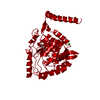
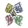
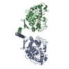
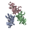
- PDB-1toh: TYROSINE HYDROXYLASE CATALYTIC AND TETRAMERIZATION DOMAINS FROM RAT -
+ Open data

- Basic information
| Entry | Database: PDB / ID: 1toh | ||||||
|---|---|---|---|---|---|---|---|
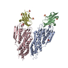
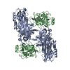
| Title | TYROSINE HYDROXYLASE CATALYTIC AND TETRAMERIZATION DOMAINS FROM RAT | ||||||
 Components Components | TYROSINE HYDROXYLASE | ||||||
 Keywords Keywords | HYDROXYLASE / NEUROTRANSMITTER BIOSYNTHESIS / NON-HEME IRON / PTERIN CO-SUBSTRATE | ||||||
| Function / homology |  Function and homology information Function and homology informationCatecholamine biosynthesis / phthalate metabolic process / tyrosine 3-monooxygenase / tyrosine 3-monooxygenase activity / dopamine biosynthetic process from tyrosine / circadian sleep/wake cycle / embryonic camera-type eye morphogenesis / epinephrine biosynthetic process / serotonin biosynthetic process / response to pyrethroid ...Catecholamine biosynthesis / phthalate metabolic process / tyrosine 3-monooxygenase / tyrosine 3-monooxygenase activity / dopamine biosynthetic process from tyrosine / circadian sleep/wake cycle / embryonic camera-type eye morphogenesis / epinephrine biosynthetic process / serotonin biosynthetic process / response to pyrethroid / aminergic neurotransmitter loading into synaptic vesicle / norepinephrine biosynthetic process / catecholamine biosynthetic process / response to curcumin / hyaloid vascular plexus regression / dopamine binding / eye photoreceptor cell development / response to ether / sphingolipid metabolic process / melanosome membrane / response to growth factor / tetrahydrobiopterin binding / response to insecticide / response to steroid hormone / synaptic transmission, dopaminergic / response to herbicide / mating behavior / response to metal ion / response to zinc ion / amino acid binding / dopamine biosynthetic process / response to corticosterone / cellular response to alkaloid / eating behavior / response to isolation stress / small molecule binding / regulation of heart contraction / response to immobilization stress / neuron projection terminus / smooth endoplasmic reticulum / response to electrical stimulus / response to light stimulus / social behavior / cellular response to manganese ion / response to salt stress / visual perception / ferric iron binding / cytoplasmic vesicle membrane / animal organ morphogenesis / response to activity / learning / response to nicotine / response to amphetamine / monooxygenase activity / cellular response to glucose stimulus / oxygen binding / locomotory behavior / ferrous iron binding / fatty acid metabolic process / response to nutrient levels / cerebral cortex development / cellular response to nicotine / cellular response to growth factor stimulus / response to peptide hormone / cellular response to xenobiotic stimulus / cognition / cytoplasmic side of plasma membrane / memory / terminal bouton / synaptic vesicle / response to estradiol / heart development / cytoplasmic vesicle / response to lipopolysaccharide / response to ethanol / perikaryon / response to hypoxia / neuron projection / response to xenobiotic stimulus / protein domain specific binding / axon / neuronal cell body / dendrite / perinuclear region of cytoplasm / enzyme binding / mitochondrion / identical protein binding / nucleus / cytoplasm Similarity search - Function | ||||||
| Biological species |  | ||||||
| Method |  X-RAY DIFFRACTION / SYNCHROTRON / MIRAS / Resolution: 2.3 Å X-RAY DIFFRACTION / SYNCHROTRON / MIRAS / Resolution: 2.3 Å | ||||||
 Authors Authors | Goodwill, K.E. / Sabatier, C. / Stevens, R.C. | ||||||
 Citation Citation | Journal: Nat.Struct.Biol. / Year: 1997 Title: Crystal structure of tyrosine hydroxylase at 2.3 A and its implications for inherited neurodegenerative diseases. Authors: Goodwill, K.E. / Sabatier, C. / Marks, C. / Raag, R. / Fitzpatrick, P.F. / Stevens, R.C. | ||||||
| History |
|
- Structure visualization
| Structure viewer | Molecule: MolmilJmol/JSmol |
|---|
- Downloads & links
-Download
| PDBx/mmCIF format | 1toh.cif.gz | 82.5 KB | Display | PDBx/mmCIF format |
|---|---|---|---|---|
| PDB format | pdb1toh.ent.gz | 61.7 KB | Display | PDB format |
| PDBx/mmJSON format | 1toh.json.gz | Tree view | PDBx/mmJSON format | |
| Others |  Other downloads Other downloads |
-Validation report
| Arichive directory | https://data.pdbj.org/pub/pdb/validation_reports/to/1tohftp://data.pdbj.org/pub/pdb/validation_reports/to/1toh | HTTPS FTP |
|---|
-Related structure data
| Similar structure data |
|---|


-Links
 PDBj
PDBj
- Assembly
| Deposited unit | 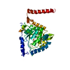
| ||||||||
|---|---|---|---|---|---|---|---|---|---|
| 1 | 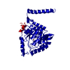
| ||||||||
| Unit cell |
|
-Components
| #1: Protein | Mass: 39274.090 Da / Num. of mol.: 1 Fragment: CATALYTIC AND TETRAMERIZATION DOMAINS, RESIDUES 156 - 498 Source method: isolated from a genetically manipulated source Details: FERRIC IRON AT THE ACTIVE SITE / Source: (gene. exp.)  |
|---|---|
| #2: Chemical | ChemComp-FE /   Mass: 55.845 Da / Num. of mol.: 1 / Source method: obtained synthetically / Formula: Fe Mass: 55.845 Da / Num. of mol.: 1 / Source method: obtained synthetically / Formula: Fe |
| #3: Water | ChemComp-HOH /  Mass: 18.015 Da / Num. of mol.: 181 / Source method: isolated from a natural source / Formula: H2O Mass: 18.015 Da / Num. of mol.: 181 / Source method: isolated from a natural source / Formula: H2O |
-Experimental details
-Experiment
| Experiment | Method: X-RAY DIFFRACTION / Number of used crystals: 1 |
|---|
- Sample preparation
| Crystal | Density Matthews: 2.59 Å3/Da / Density % sol: 46 % Description: THREE DERIVATIVES WITH ANOMALOUS DATE WERE USED. | |||||||||||||||||||||||||||||||||||||||||||||||||||||||
|---|---|---|---|---|---|---|---|---|---|---|---|---|---|---|---|---|---|---|---|---|---|---|---|---|---|---|---|---|---|---|---|---|---|---|---|---|---|---|---|---|---|---|---|---|---|---|---|---|---|---|---|---|---|---|---|---|
| Crystal grow | Temperature: 277 K / pH: 7.9 Details: 1.2 M AMMONIUM SULFATE 3.2% PEG 200 5% GLYCEROL 2 MM DTT 80 MM TRIS, PH 7.9 CRYSTALS GROWN AT 4 DEGREES C, temperature 277K | |||||||||||||||||||||||||||||||||||||||||||||||||||||||
| Crystal grow | *PLUS Temperature: 4 K / Method: microdialysis | |||||||||||||||||||||||||||||||||||||||||||||||||||||||
| Components of the solutions | *PLUS
|
-Data collection
| Diffraction | Mean temperature: 100 K |
|---|---|
| Diffraction source | Source: SYNCHROTRON / Site: SSRL  / Beamline: BL7-1 / Wavelength: 1.08 / Beamline: BL7-1 / Wavelength: 1.08 |
| Detector | Type: MARRESEARCH / Detector: IMAGE PLATE / Date: Jun 1, 1996 / Details: MIRRORS |
| Radiation | Monochromator: DOUBLE CRYSTAL SI(111) / Monochromatic (M) / Laue (L): M / Scattering type: x-ray |
| Radiation wavelength | Wavelength: 1.08 Å / Relative weight: 1 |
| Reflection | Resolution: 2.3→30 Å / Num. obs: 18068 / % possible obs: 98 % / Observed criterion σ(I): 2 / Redundancy: 7 % / Biso Wilson estimate: 54.1 Å2 / Rmerge(I) obs: 0.042 / Rsym value: 0.042 / Net I/σ(I): 21.3 |
| Reflection shell | Resolution: 2.3→2.38 Å / Redundancy: 5.5 % / Rmerge(I) obs: 0.187 / Mean I/σ(I) obs: 5 / Rsym value: 0.187 / % possible all: 92 |
| Reflection | *PLUS Num. measured all: 126857 |
| Reflection shell | *PLUS % possible obs: 92 % |
- Processing
| Software |
| ||||||||||||||||||||||||||||||||||||||||||||||||||||||||||||||||||||||||||||||||
|---|---|---|---|---|---|---|---|---|---|---|---|---|---|---|---|---|---|---|---|---|---|---|---|---|---|---|---|---|---|---|---|---|---|---|---|---|---|---|---|---|---|---|---|---|---|---|---|---|---|---|---|---|---|---|---|---|---|---|---|---|---|---|---|---|---|---|---|---|---|---|---|---|---|---|---|---|---|---|---|---|---|
| Refinement | Method to determine structure: MIRAS / Resolution: 2.3→37 Å / Rfactor Rfree error: 0.008 / Data cutoff high absF: 2194380.94 / Data cutoff low absF: 0 / Isotropic thermal model: RESTRAINED / Cross valid method: THROUGHOUT / σ(F): 2 / Stereochemistry target values: MAXIMUM LIKELIHOOD FUNCTION
| ||||||||||||||||||||||||||||||||||||||||||||||||||||||||||||||||||||||||||||||||
| Solvent computation | Bsol: 42.71 Å2 / ksol: 0.341 e/Å3 | ||||||||||||||||||||||||||||||||||||||||||||||||||||||||||||||||||||||||||||||||
| Displacement parameters | Biso mean: 46.4 Å2
| ||||||||||||||||||||||||||||||||||||||||||||||||||||||||||||||||||||||||||||||||
| Refine analyze |
| ||||||||||||||||||||||||||||||||||||||||||||||||||||||||||||||||||||||||||||||||
| Refinement step | Cycle: LAST / Resolution: 2.3→37 Å
| ||||||||||||||||||||||||||||||||||||||||||||||||||||||||||||||||||||||||||||||||
| Refine LS restraints |
| ||||||||||||||||||||||||||||||||||||||||||||||||||||||||||||||||||||||||||||||||
| LS refinement shell | Resolution: 2.3→2.44 Å / Rfactor Rfree error: 0.024 / Total num. of bins used: 6
| ||||||||||||||||||||||||||||||||||||||||||||||||||||||||||||||||||||||||||||||||
| Xplor file |
| ||||||||||||||||||||||||||||||||||||||||||||||||||||||||||||||||||||||||||||||||
| Software | *PLUS Name: CNS / Version: 0.3 / Classification: refinement | ||||||||||||||||||||||||||||||||||||||||||||||||||||||||||||||||||||||||||||||||
| Refinement | *PLUS | ||||||||||||||||||||||||||||||||||||||||||||||||||||||||||||||||||||||||||||||||
| Solvent computation | *PLUS | ||||||||||||||||||||||||||||||||||||||||||||||||||||||||||||||||||||||||||||||||
| Displacement parameters | *PLUS | ||||||||||||||||||||||||||||||||||||||||||||||||||||||||||||||||||||||||||||||||
| Refine LS restraints | *PLUS
| ||||||||||||||||||||||||||||||||||||||||||||||||||||||||||||||||||||||||||||||||
| LS refinement shell | *PLUS Rfactor obs: 0.265 |
