 Movie
Movie Controller


+ Open data

- Basic information
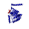
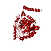
| Entry | Database: PDB / ID: 2pah | ||||||
|---|---|---|---|---|---|---|---|
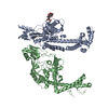
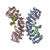
| Title | TETRAMERIC HUMAN PHENYLALANINE HYDROXYLASE | ||||||
 Components Components | PROTEIN (PHENYLALANINE HYDROXYLASE) | ||||||
 Keywords Keywords | HYDROXYLASE / PHENYLKETONURIA | ||||||
| Function / homology |  Function and homology information Function and homology informationPhenylketonuria / phenylalanine 4-monooxygenase / Phenylalanine metabolism / phenylalanine 4-monooxygenase activity / L-tyrosine biosynthetic process / catecholamine biosynthetic process / L-phenylalanine catabolic process / amino acid biosynthetic process / iron ion binding / cytosol Similarity search - Function | ||||||
| Biological species |  Homo sapiens (human) Homo sapiens (human) | ||||||
| Method |  X-RAY DIFFRACTION / SYNCHROTRON / MOLECULAR REPLACEMENT / Resolution: 3.1 Å X-RAY DIFFRACTION / SYNCHROTRON / MOLECULAR REPLACEMENT / Resolution: 3.1 Å | ||||||
 Authors Authors | Stevens, R.C. / Fusetti, F. / Erlandsen, H. | ||||||
 Citation Citation | Journal: J.Biol.Chem. / Year: 1998 Title: Structure of tetrameric human phenylalanine hydroxylase and its implications for phenylketonuria. Authors: Fusetti, F. / Erlandsen, H. / Flatmark, T. / Stevens, R.C. | ||||||
| History |
|
- Structure visualization
| Structure viewer | Molecule: MolmilJmol/JSmol |
|---|
- Downloads & links
-Download
| PDBx/mmCIF format | 2pah.cif.gz | 138.2 KB | Display | PDBx/mmCIF format |
|---|---|---|---|---|
| PDB format | pdb2pah.ent.gz | 108.8 KB | Display | PDB format |
| PDBx/mmJSON format | 2pah.json.gz | Tree view | PDBx/mmJSON format | |
| Others |  Other downloads Other downloads |
-Validation report
| Arichive directory | https://data.pdbj.org/pub/pdb/validation_reports/pa/2pahftp://data.pdbj.org/pub/pdb/validation_reports/pa/2pah | HTTPS FTP |
|---|
-Related structure data
| Related structure data |  1tohS S: Starting model for refinement |
|---|---|
| Similar structure data |


-Links
 PDBj
PDBj

- Assembly
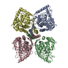
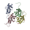
| Deposited unit | 
| ||||||||
|---|---|---|---|---|---|---|---|---|---|
| 1 | 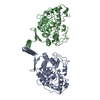
| ||||||||
| Unit cell |
|
-Components
| #1: Protein | Mass: 38641.926 Da / Num. of mol.: 2 / Fragment: RESIDUES 118-452 Source method: isolated from a genetically manipulated source Details: ACTIVE SITE IRON ATOM FE / Source: (gene. exp.) Homo sapiens (human) / Plasmid: PET-3A-D / Species (production host): Escherichia coli / Production host:  #2: Chemical |   Mass: 55.845 Da / Num. of mol.: 2 / Source method: obtained synthetically / Formula: Fe Mass: 55.845 Da / Num. of mol.: 2 / Source method: obtained synthetically / Formula: Fe |
|---|
-Experimental details
-Experiment
| Experiment | Method: X-RAY DIFFRACTION / Number of used crystals: 1 |
|---|
- Sample preparation
| Crystal | Density Matthews: 3.36 Å3/Da / Density % sol: 60 % | ||||||||||||||||||||||||||||||
|---|---|---|---|---|---|---|---|---|---|---|---|---|---|---|---|---|---|---|---|---|---|---|---|---|---|---|---|---|---|---|---|
| Crystal grow | pH: 7 Details: 0.6 M MGSO4, 0.2 M NAK TARTRATE, 10% PEG-4000, 0.1 M BIS-TRIS PROPANE (PH 7.0) | ||||||||||||||||||||||||||||||
| Crystal | *PLUS Density % sol: 60 % | ||||||||||||||||||||||||||||||
| Crystal grow | *PLUS Method: vapor diffusion, hanging drop | ||||||||||||||||||||||||||||||
| Components of the solutions | *PLUS
|
-Data collection
| Diffraction | Mean temperature: 100 K |
|---|---|
| Diffraction source | Source: SYNCHROTRON / Site: SSRL  / Beamline: BL7-1 / Wavelength: 1.08 / Beamline: BL7-1 / Wavelength: 1.08 |
| Detector | Type: MARRESEARCH / Detector: IMAGE PLATE / Date: Jul 1, 1997 / Details: MIRRORS |
| Radiation | Protocol: SINGLE WAVELENGTH / Monochromatic (M) / Laue (L): M / Scattering type: x-ray |
| Radiation wavelength | Wavelength: 1.08 Å / Relative weight: 1 |
| Reflection | Resolution: 3.1→20 Å / Num. obs: 120922 / % possible obs: 99 % / Observed criterion σ(I): 0 / Redundancy: 6.5 % / Biso Wilson estimate: 33 Å2 / Rmerge(I) obs: 0.075 / Rsym value: 0.075 / Net I/σ(I): 10 |
| Reflection shell | Resolution: 3.1→3.2 Å / Redundancy: 2 % / Rmerge(I) obs: 0.25 / Mean I/σ(I) obs: 2 / Rsym value: 0.25 / % possible all: 98 |
| Reflection | *PLUS Num. obs: 18446 / Num. measured all: 120922 |
- Processing
| Software |
| ||||||||||||||||||||||||||||||||||||||||||||||||||||||||||||
|---|---|---|---|---|---|---|---|---|---|---|---|---|---|---|---|---|---|---|---|---|---|---|---|---|---|---|---|---|---|---|---|---|---|---|---|---|---|---|---|---|---|---|---|---|---|---|---|---|---|---|---|---|---|---|---|---|---|---|---|---|---|
| Refinement | Method to determine structure: MOLECULAR REPLACEMENT Starting model: 1TOH Resolution: 3.1→20 Å / Data cutoff high absF: 100000000 / Data cutoff low absF: 0.001 / Cross valid method: THROUGHOUT / σ(F): 0
| ||||||||||||||||||||||||||||||||||||||||||||||||||||||||||||
| Displacement parameters | Biso mean: 33 Å2
| ||||||||||||||||||||||||||||||||||||||||||||||||||||||||||||
| Refinement step | Cycle: LAST / Resolution: 3.1→20 Å
| ||||||||||||||||||||||||||||||||||||||||||||||||||||||||||||
| Refine LS restraints |
| ||||||||||||||||||||||||||||||||||||||||||||||||||||||||||||
| Refine LS restraints NCS | NCS model details: RESTRAINTS | ||||||||||||||||||||||||||||||||||||||||||||||||||||||||||||
| LS refinement shell | Resolution: 3.1→3.2 Å / Total num. of bins used: 6 /
| ||||||||||||||||||||||||||||||||||||||||||||||||||||||||||||
| Xplor file |
| ||||||||||||||||||||||||||||||||||||||||||||||||||||||||||||
| Software | *PLUS Name: X-PLOR / Version: 3.8 / Classification: refinement | ||||||||||||||||||||||||||||||||||||||||||||||||||||||||||||
| Refinement | *PLUS Highest resolution: 3.1 Å / Lowest resolution: 20 Å / σ(F): 0 / % reflection Rfree: 10 % | ||||||||||||||||||||||||||||||||||||||||||||||||||||||||||||
| Solvent computation | *PLUS | ||||||||||||||||||||||||||||||||||||||||||||||||||||||||||||
| Displacement parameters | *PLUS Biso mean: 33 Å2 | ||||||||||||||||||||||||||||||||||||||||||||||||||||||||||||
| Refine LS restraints | *PLUS Type: x_angle_deg / Dev ideal: 1.7 | ||||||||||||||||||||||||||||||||||||||||||||||||||||||||||||
| LS refinement shell | *PLUS Highest resolution: 3.1 Å / Lowest resolution: 3.2 Å / Rfactor Rfree: 0.326 / % reflection Rfree: 10 % / Rfactor Rwork: 0.251 |
