[English] 日本語
 Yorodumi
- PDB-6wwz: Cryo-EM structure of the human chemokine receptor CCR6 in complex... -
+ Open data
- Basic information
| Entry | Database: PDB / ID: 6wwz | ||||||
|---|---|---|---|---|---|---|---|
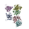
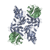
| Title | Cryo-EM structure of the human chemokine receptor CCR6 in complex with CCL20 and a Go protein | ||||||
 Components Components |
| ||||||
 Keywords Keywords | MEMBRANE PROTEIN / GPCR / Chemokine / Chemokine receptor / Complex | ||||||
| Function / homology |  Function and homology information Function and homology informationisotype switching to IgA isotypes / DN3 thymocyte differentiation / thymocyte migration / positive regulation of flagellated sperm motility involved in capacitation / CCR6 chemokine receptor binding / Beta defensins / sperm principal piece / positive regulation of dendritic cell chemotaxis / regulation of T cell migration / lymphocyte migration ...isotype switching to IgA isotypes / DN3 thymocyte differentiation / thymocyte migration / positive regulation of flagellated sperm motility involved in capacitation / CCR6 chemokine receptor binding / Beta defensins / sperm principal piece / positive regulation of dendritic cell chemotaxis / regulation of T cell migration / lymphocyte migration / DN2 thymocyte differentiation / chemokine receptor activity / leukocyte migration involved in inflammatory response / C-C chemokine receptor activity / mu-type opioid receptor binding / C-C chemokine binding / corticotropin-releasing hormone receptor 1 binding / G-protein activation / Activation of the phototransduction cascade / Glucagon-type ligand receptors / Thromboxane signalling through TP receptor / Sensory perception of sweet, bitter, and umami (glutamate) taste / G beta:gamma signalling through PI3Kgamma / G beta:gamma signalling through CDC42 / Cooperation of PDCL (PhLP1) and TRiC/CCT in G-protein beta folding / Activation of G protein gated Potassium channels / Inhibition of voltage gated Ca2+ channels via Gbeta/gamma subunits / Ca2+ pathway / G alpha (z) signalling events / High laminar flow shear stress activates signaling by PIEZO1 and PECAM1:CDH5:KDR in endothelial cells / Glucagon-like Peptide-1 (GLP1) regulates insulin secretion / Vasopressin regulates renal water homeostasis via Aquaporins / Adrenaline,noradrenaline inhibits insulin secretion / ADP signalling through P2Y purinoceptor 12 / G alpha (q) signalling events / chemokine activity / vesicle docking involved in exocytosis / G alpha (i) signalling events / Chemokine receptors bind chemokines / Thrombin signalling through proteinase activated receptors (PARs) / dendritic cell chemotaxis / photoreceptor outer segment membrane / G protein-coupled dopamine receptor signaling pathway / spectrin binding / sperm plasma membrane / regulation of heart contraction / alkylglycerophosphoethanolamine phosphodiesterase activity / parallel fiber to Purkinje cell synapse / Interleukin-10 signaling / humoral immune response / photoreceptor outer segment / cellular defense response / sperm flagellum / positive regulation of T cell migration / postsynaptic modulation of chemical synaptic transmission / T cell migration / sperm midpiece / cardiac muscle cell apoptotic process / photoreceptor inner segment / adenylate cyclase regulator activity / G protein-coupled serotonin receptor binding / adenylate cyclase-inhibiting serotonin receptor signaling pathway / muscle contraction / cell chemotaxis / locomotory behavior / calcium-mediated signaling / electron transport chain / negative regulation of insulin secretion / GABA-ergic synapse / adenylate cyclase-modulating G protein-coupled receptor signaling pathway / G-protein beta/gamma-subunit complex binding / chemotaxis / G beta:gamma signalling through PLC beta / Presynaptic function of Kainate receptors / Thromboxane signalling through TP receptor / Activation of G protein gated Potassium channels / Inhibition of voltage gated Ca2+ channels via Gbeta/gamma subunits / G-protein activation / Prostacyclin signalling through prostacyclin receptor / G beta:gamma signalling through CDC42 / Glucagon signaling in metabolic regulation / G beta:gamma signalling through BTK / ADP signalling through P2Y purinoceptor 12 / Glucagon-type ligand receptors / Adrenaline,noradrenaline inhibits insulin secretion / Vasopressin regulates renal water homeostasis via Aquaporins / Glucagon-like Peptide-1 (GLP1) regulates insulin secretion / G alpha (z) signalling events / cellular response to catecholamine stimulus / ADP signalling through P2Y purinoceptor 1 / ADORA2B mediated anti-inflammatory cytokines production / G beta:gamma signalling through PI3Kgamma / Cooperation of PDCL (PhLP1) and TRiC/CCT in G-protein beta folding / adenylate cyclase-activating dopamine receptor signaling pathway / cell-cell signaling / GPER1 signaling / G-protein beta-subunit binding / cellular response to prostaglandin E stimulus / heterotrimeric G-protein complex / signaling receptor activity Similarity search - Function | ||||||
| Biological species |   Homo sapiens (human) Homo sapiens (human) | ||||||
| Method | ELECTRON MICROSCOPY / single particle reconstruction / cryo EM / Resolution: 3.34 Å | ||||||
 Authors Authors | Wasilko, D.J. / Johnson, Z.L. / Ammirati, M. / Han, S. / Wu, H. | ||||||
 Citation Citation |  Journal: Nat Commun / Year: 2020 Journal: Nat Commun / Year: 2020Title: Structural basis for chemokine receptor CCR6 activation by the endogenous protein ligand CCL20. Authors: David Jonathan Wasilko / Zachary Lee Johnson / Mark Ammirati / Ye Che / Matthew C Griffor / Seungil Han / Huixian Wu /  Abstract: Chemokines are important protein-signaling molecules that regulate various immune responses by activating chemokine receptors which belong to the G protein-coupled receptor (GPCR) superfamily. ...Chemokines are important protein-signaling molecules that regulate various immune responses by activating chemokine receptors which belong to the G protein-coupled receptor (GPCR) superfamily. Despite the substantial progression of our structural understanding of GPCR activation by small molecule and peptide agonists, the molecular mechanism of GPCR activation by protein agonists remains unclear. Here, we present a 3.3-Å cryo-electron microscopy structure of the human chemokine receptor CCR6 bound to its endogenous ligand CCL20 and an engineered Go. CCL20 binds in a shallow extracellular pocket, making limited contact with the core 7-transmembrane (TM) bundle. The structure suggests that this mode of binding induces allosterically a rearrangement of a noncanonical toggle switch and the opening of the intracellular crevice for G protein coupling. Our results demonstrate that GPCR activation by a protein agonist does not always require substantial interactions between ligand and the 7TM core region. | ||||||
| History |
|
- Structure visualization
| Movie |
 Movie viewer Movie viewer |
|---|---|
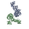
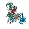
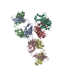
| Structure viewer | Molecule:  Molmil Molmil Jmol/JSmol Jmol/JSmol |
- Downloads & links
- Download
| PDBx/mmCIF format |  6wwz.cif.gz 6wwz.cif.gz | 256.5 KB | Display |  PDBx/mmCIF format PDBx/mmCIF format |
|---|---|---|---|---|
| PDB format |  pdb6wwz.ent.gz pdb6wwz.ent.gz | 196.5 KB | Display |  PDB format PDB format |
| PDBx/mmJSON format |  6wwz.json.gz 6wwz.json.gz | Tree view |  PDBx/mmJSON format PDBx/mmJSON format | |
| Others |  Other downloads Other downloads |
-Validation report
| Summary document |  6wwz_validation.pdf.gz 6wwz_validation.pdf.gz | 874 KB | Display |  wwPDB validaton report wwPDB validaton report |
|---|---|---|---|---|
| Full document |  6wwz_full_validation.pdf.gz 6wwz_full_validation.pdf.gz | 875.3 KB | Display | |
| Data in XML |  6wwz_validation.xml.gz 6wwz_validation.xml.gz | 41.2 KB | Display | |
| Data in CIF |  6wwz_validation.cif.gz 6wwz_validation.cif.gz | 63.8 KB | Display | |
| Arichive directory |  https://data.pdbj.org/pub/pdb/validation_reports/ww/6wwz https://data.pdbj.org/pub/pdb/validation_reports/ww/6wwz ftp://data.pdbj.org/pub/pdb/validation_reports/ww/6wwz ftp://data.pdbj.org/pub/pdb/validation_reports/ww/6wwz | HTTPS FTP |
-Related structure data
| Related structure data |  21950MC M: map data used to model this data C: citing same article ( |
|---|---|
| Similar structure data |
- Links
Links
- Assembly
| Deposited unit | 
|
|---|---|
| 1 |
|
- Components
-Guanine nucleotide-binding protein ... , 3 types, 3 molecules BYA
| #1: Protein | Mass: 38744.371 Da / Num. of mol.: 1 Source method: isolated from a genetically manipulated source Source: (gene. exp.)   |
|---|---|
| #3: Protein | Mass: 7563.750 Da / Num. of mol.: 1 Source method: isolated from a genetically manipulated source Source: (gene. exp.)   |
| #4: Protein | Mass: 28195.869 Da / Num. of mol.: 1 Source method: isolated from a genetically manipulated source Source: (gene. exp.)  Homo sapiens (human) / Gene: GNAO1 / Production host: Homo sapiens (human) / Gene: GNAO1 / Production host:  |
-Protein , 2 types, 2 molecules CR
| #2: Protein | Mass: 8043.568 Da / Num. of mol.: 1 Source method: isolated from a genetically manipulated source Source: (gene. exp.)  Homo sapiens (human) / Gene: CCL20, LARC, MIP3A, SCYA20 / Production host: Homo sapiens (human) / Gene: CCL20, LARC, MIP3A, SCYA20 / Production host:  |
|---|---|
| #6: Protein | Mass: 60064.574 Da / Num. of mol.: 1 Source method: isolated from a genetically manipulated source Source: (gene. exp.)  Homo sapiens (human) / Gene: CCR6, CKRL3, CMKBR6, GPR29, STRL22 / Production host: Homo sapiens (human) / Gene: CCR6, CKRL3, CMKBR6, GPR29, STRL22 / Production host:  |
-Antibody , 1 types, 1 molecules S
| #5: Antibody | Mass: 30895.527 Da / Num. of mol.: 1 Source method: isolated from a genetically manipulated source Source: (gene. exp.)   |
|---|
-Details
| Has protein modification | Y |
|---|
-Experimental details
-Experiment
| Experiment | Method: ELECTRON MICROSCOPY |
|---|---|
| EM experiment | Aggregation state: PARTICLE / 3D reconstruction method: single particle reconstruction |
- Sample preparation
| Component |
| |||||||||||||||||||||||||||||||||||
|---|---|---|---|---|---|---|---|---|---|---|---|---|---|---|---|---|---|---|---|---|---|---|---|---|---|---|---|---|---|---|---|---|---|---|---|---|
| Molecular weight | Experimental value: NO | |||||||||||||||||||||||||||||||||||
| Source (natural) |
| |||||||||||||||||||||||||||||||||||
| Source (recombinant) |
| |||||||||||||||||||||||||||||||||||
| Buffer solution | pH: 7.5 | |||||||||||||||||||||||||||||||||||
| Buffer component |
| |||||||||||||||||||||||||||||||||||
| Specimen | Conc.: 6.8 mg/ml / Embedding applied: NO / Shadowing applied: NO / Staining applied: NO / Vitrification applied: YES | |||||||||||||||||||||||||||||||||||
| Specimen support | Grid material: GOLD / Grid mesh size: 300 divisions/in. / Grid type: Quantifoil R1.2/1.3 | |||||||||||||||||||||||||||||||||||
| Vitrification | Instrument: FEI VITROBOT MARK IV / Cryogen name: ETHANE / Humidity: 100 % / Chamber temperature: 277 K |
- Electron microscopy imaging
| Experimental equipment |  Model: Titan Krios / Image courtesy: FEI Company |
|---|---|
| Microscopy | Model: FEI TITAN KRIOS |
| Electron gun | Electron source:  FIELD EMISSION GUN / Accelerating voltage: 300 kV / Illumination mode: FLOOD BEAM FIELD EMISSION GUN / Accelerating voltage: 300 kV / Illumination mode: FLOOD BEAM |
| Electron lens | Mode: BRIGHT FIELD / Nominal magnification: 165000 X / Nominal defocus max: 2000 nm / Nominal defocus min: 600 nm / Cs: 2.7 mm / C2 aperture diameter: 100 µm |
| Specimen holder | Cryogen: NITROGEN / Specimen holder model: FEI TITAN KRIOS AUTOGRID HOLDER / Temperature (max): 100 K / Temperature (min): 80 K |
| Image recording | Average exposure time: 8 sec. / Electron dose: 81 e/Å2 / Detector mode: SUPER-RESOLUTION / Film or detector model: GATAN K2 SUMMIT (4k x 4k) / Num. of grids imaged: 1 / Num. of real images: 5303 |
| EM imaging optics | Energyfilter name: GIF Quantum LS / Energyfilter slit width: 20 eV |
| Image scans | Width: 3838 / Height: 3710 / Movie frames/image: 50 / Used frames/image: 1-50 |
- Processing
| Software | Name: REFMAC / Version: 5.8.0258 / Classification: refinement | ||||||||||||||||||||||||||||||||||||||||||||||||||||||||||||||||||||||||||||||||||||||||||||||||||||||||||||
|---|---|---|---|---|---|---|---|---|---|---|---|---|---|---|---|---|---|---|---|---|---|---|---|---|---|---|---|---|---|---|---|---|---|---|---|---|---|---|---|---|---|---|---|---|---|---|---|---|---|---|---|---|---|---|---|---|---|---|---|---|---|---|---|---|---|---|---|---|---|---|---|---|---|---|---|---|---|---|---|---|---|---|---|---|---|---|---|---|---|---|---|---|---|---|---|---|---|---|---|---|---|---|---|---|---|---|---|---|---|
| EM software |
| ||||||||||||||||||||||||||||||||||||||||||||||||||||||||||||||||||||||||||||||||||||||||||||||||||||||||||||
| CTF correction | Type: PHASE FLIPPING AND AMPLITUDE CORRECTION | ||||||||||||||||||||||||||||||||||||||||||||||||||||||||||||||||||||||||||||||||||||||||||||||||||||||||||||
| Particle selection | Num. of particles selected: 1000382 | ||||||||||||||||||||||||||||||||||||||||||||||||||||||||||||||||||||||||||||||||||||||||||||||||||||||||||||
| Symmetry | Point symmetry: C1 (asymmetric) | ||||||||||||||||||||||||||||||||||||||||||||||||||||||||||||||||||||||||||||||||||||||||||||||||||||||||||||
| 3D reconstruction | Resolution: 3.34 Å / Resolution method: FSC 0.143 CUT-OFF / Num. of particles: 230450 / Num. of class averages: 1 / Symmetry type: POINT | ||||||||||||||||||||||||||||||||||||||||||||||||||||||||||||||||||||||||||||||||||||||||||||||||||||||||||||
| Atomic model building |
| ||||||||||||||||||||||||||||||||||||||||||||||||||||||||||||||||||||||||||||||||||||||||||||||||||||||||||||
| Atomic model building | Source name: PDB / Type: experimental model
| ||||||||||||||||||||||||||||||||||||||||||||||||||||||||||||||||||||||||||||||||||||||||||||||||||||||||||||
| Refinement | Resolution: 3.34→154 Å / Cor.coef. Fo:Fc: 0.86 / SU B: 109.675 / SU ML: 2.255 / ESU R: 0.432 Stereochemistry target values: MAXIMUM LIKELIHOOD WITH PHASES Details: HYDROGENS HAVE BEEN ADDED IN THE RIDING POSITIONS
| ||||||||||||||||||||||||||||||||||||||||||||||||||||||||||||||||||||||||||||||||||||||||||||||||||||||||||||
| Solvent computation | Ion probe radii: 0.8 Å / Shrinkage radii: 0.8 Å / VDW probe radii: 1.2 Å / Solvent model: MASK | ||||||||||||||||||||||||||||||||||||||||||||||||||||||||||||||||||||||||||||||||||||||||||||||||||||||||||||
| Displacement parameters | Biso mean: 107.024 Å2
|
 Movie
Movie Controller



 PDBj
PDBj


