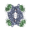
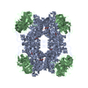
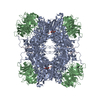
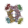
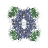
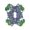
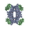
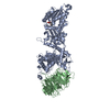
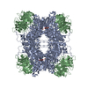
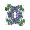
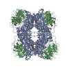
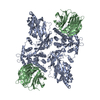
Entry Database : PDB / ID : 5fa5Title Crystal Structure of PRMT5:MEP50 in complex with MTA and H4 peptide Histone H4 Methylosome protein 50 Protein arginine N-methyltransferase 5 Keywords / / Function / homology Function Domain/homology Component
/ / / / / / / / / / / / / / / / / / / / / / / / / / / / / / / / / / / / / / / / / / / / / / / / / / / / / / / / / / / / / / / / / / / / / / / / / / / / / / / / / / / / / / / / / / / / / / / / / / / / / / / / / / / / / / / / / / / / / / / / / / / / / / / / / / / / / / / / / / / / / / / / / / / / / / / / / / / / / / / Biological species Homo sapiens (human)Method / / / / Resolution : 2.34 Å Authors Sprague, E.R. / McNamara, J.T. Journal : Science / Year : 2016Title : Disordered methionine metabolism in MTAP/CDKN2A-deleted cancers leads to dependence on PRMT5.Authors: Mavrakis, K.J. / McDonald, E.R. / Schlabach, M.R. / Billy, E. / Hoffman, G.R. / deWeck, A. / Ruddy, D.A. / Venkatesan, K. / Yu, J. / McAllister, G. / Stump, M. / deBeaumont, R. / Ho, S. / ... Authors : Mavrakis, K.J. / McDonald, E.R. / Schlabach, M.R. / Billy, E. / Hoffman, G.R. / deWeck, A. / Ruddy, D.A. / Venkatesan, K. / Yu, J. / McAllister, G. / Stump, M. / deBeaumont, R. / Ho, S. / Yue, Y. / Liu, Y. / Yan-Neale, Y. / Yang, G. / Lin, F. / Yin, H. / Gao, H. / Kipp, D.R. / Zhao, S. / McNamara, J.T. / Sprague, E.R. / Zheng, B. / Lin, Y. / Cho, Y.S. / Gu, J. / Crawford, K. / Ciccone, D. / Vitari, A.C. / Lai, A. / Capka, V. / Hurov, K. / Porter, J.A. / Tallarico, J. / Mickanin, C. / Lees, E. / Pagliarini, R. / Keen, N. / Schmelzle, T. / Hofmann, F. / Stegmeier, F. / Sellers, W.R. History Deposition Dec 10, 2015 Deposition site / Processing site Revision 1.0 Feb 24, 2016 Provider / Type Revision 1.1 Mar 9, 2016 Group Revision 1.2 Mar 23, 2016 Group Revision 1.3 Sep 27, 2023 Group Data collection / Database references ... Data collection / Database references / Derived calculations / Refinement description Category chem_comp_atom / chem_comp_bond ... chem_comp_atom / chem_comp_bond / citation / database_2 / pdbx_initial_refinement_model / pdbx_struct_oper_list Item _citation.journal_id_CSD / _database_2.pdbx_DOI ... _citation.journal_id_CSD / _database_2.pdbx_DOI / _database_2.pdbx_database_accession / _pdbx_struct_oper_list.symmetry_operation
Show all Show less
 Movie
Movie Controller

 Yorodumi
 Open data
 Basic information
 Components
 Keywords
 Function and homology information
 Homo sapiens (human)
 X-RAY DIFFRACTION /
  Authors
 Citation
 Structure visualization
 Downloads & links
 Other downloads
 PDBj
PDBj


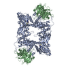
 Assembly
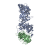

 Spodoptera frugiperda (fall armyworm) / Strain (production host): Sf21
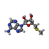
 Mass: 297.334 Da / Num. of mol.: 1 / Source method: obtained synthetically / Formula: C11H15N5O3S
 Mass: 18.015 Da / Num. of mol.: 168 / Source method: isolated from a natural source / Formula: H2O
 Sample preparation
 / Beamline: 17-ID / Wavelength: 1 Å
 Processing