 Movie
Movie Controller

[English] 日本語
 Yorodumi
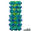
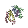
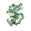
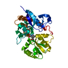
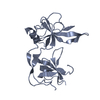
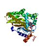
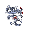
- PDB-6vci: Lipophilic envelope-spanning tunnel protein (LetB), domains MCE2-MCE3 -
+ Open data

- Basic information
| Entry | Database: PDB / ID: 6vci | ||||||||||||||||||
|---|---|---|---|---|---|---|---|---|---|---|---|---|---|---|---|---|---|---|---|
| Title | Lipophilic envelope-spanning tunnel protein (LetB), domains MCE2-MCE3 | ||||||||||||||||||
 Components Components | Lipophilic envelope-spanning tunnel protein LetB | ||||||||||||||||||
 Keywords Keywords | LIPID TRANSPORT / bacterial cell envelope / MCE / YebT / LetB | ||||||||||||||||||
| Function / homology | : / Mce/MlaD / MlaD protein / intermembrane lipid transfer / membrane organization / outer membrane-bounded periplasmic space / identical protein binding / plasma membrane / Lipophilic envelope-spanning tunnel protein B Function and homology information Function and homology information | ||||||||||||||||||
| Biological species |  | ||||||||||||||||||
| Method |  X-RAY DIFFRACTION / SYNCHROTRON / MOLECULAR REPLACEMENT / Resolution: 2.15 Å X-RAY DIFFRACTION / SYNCHROTRON / MOLECULAR REPLACEMENT / Resolution: 2.15 Å | ||||||||||||||||||
 Authors Authors | Isom, G.L. / Coudray, N. / MacRae, M.R. / McManus, C.T. / Ekiert, D.C. / Bhabha, G. | ||||||||||||||||||
| Funding support |  United States, 5items United States, 5items
| ||||||||||||||||||
 Citation Citation | Journal: Cell / Year: 2020 Title: LetB Structure Reveals a Tunnel for Lipid Transport across the Bacterial Envelope. Authors: Georgia L Isom / Nicolas Coudray / Mark R MacRae / Collin T McManus / Damian C Ekiert / Gira Bhabha / Abstract: Gram-negative bacteria are surrounded by an outer membrane composed of phospholipids and lipopolysaccharide, which acts as a barrier and contributes to antibiotic resistance. The systems that mediate ...Gram-negative bacteria are surrounded by an outer membrane composed of phospholipids and lipopolysaccharide, which acts as a barrier and contributes to antibiotic resistance. The systems that mediate phospholipid trafficking across the periplasm, such as MCE (Mammalian Cell Entry) transporters, have not been well characterized. Our ~3.5 Å cryo-EM structure of the E. coli MCE protein LetB reveals an ~0.6 megadalton complex that consists of seven stacked rings, with a central hydrophobic tunnel sufficiently long to span the periplasm. Lipids bind inside the tunnel, suggesting that it functions as a pathway for lipid transport. Cryo-EM structures in the open and closed states reveal a dynamic tunnel lining, with implications for gating or substrate translocation. Our results support a model in which LetB establishes a physical link between the two membranes and creates a hydrophobic pathway for the translocation of lipids across the periplasm. | ||||||||||||||||||
| History |
|
- Structure visualization
| Structure viewer | Molecule: MolmilJmol/JSmol |
|---|
- Downloads & links
-Download
| PDBx/mmCIF format | 6vci.cif.gz | 179.7 KB | Display | PDBx/mmCIF format |
|---|---|---|---|---|
| PDB format | pdb6vci.ent.gz | 141.6 KB | Display | PDB format |
| PDBx/mmJSON format | 6vci.json.gz | Tree view | PDBx/mmJSON format | |
| Others |  Other downloads Other downloads |
-Validation report
| Arichive directory | https://data.pdbj.org/pub/pdb/validation_reports/vc/6vciftp://data.pdbj.org/pub/pdb/validation_reports/vc/6vci | HTTPS FTP |
|---|
-Related structure data
| Related structure data |  6v0cC  6v0dC  6v0eC  6v0fSC 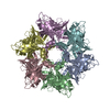 6v0gC  6v0hC  6v0iC  6v0jC S: Starting model for refinement C: citing same article ( |
|---|---|
| Similar structure data |


-Links
 PDBj
PDBj- Assembly
| Deposited unit | 
| ||||||||||||
|---|---|---|---|---|---|---|---|---|---|---|---|---|---|
| 1 | 
| ||||||||||||
| 2 | 
| ||||||||||||
| Unit cell |
|
-Components
| #1: Protein | Mass: 25419.844 Da / Num. of mol.: 2 Source method: isolated from a genetically manipulated source Source: (gene. exp.) Strain: K12 / Gene: yebT, b1834, JW1823 / Production host: #2: Water | ChemComp-HOH / |  Mass: 18.015 Da / Num. of mol.: 48 / Source method: isolated from a natural source / Formula: H2O Mass: 18.015 Da / Num. of mol.: 48 / Source method: isolated from a natural source / Formula: H2O |
|---|
-Experimental details
-Experiment
| Experiment | Method: X-RAY DIFFRACTION / Number of used crystals: 1 |
|---|
- Sample preparation
| Crystal | Density Matthews: 2.68 Å3/Da / Density % sol: 54.07 % |
|---|---|
| Crystal grow | Temperature: 291.15 K / Method: vapor diffusion, sitting drop / pH: 8.5 Details: Crystals grew from drops consisting of 100 nL protein plus 100 nL of a reservoir solution consisting of 0.1 M Tris pH 8.5, 20% w/v PEG 1K, and were cryoprotected by supplementing the ...Details: Crystals grew from drops consisting of 100 nL protein plus 100 nL of a reservoir solution consisting of 0.1 M Tris pH 8.5, 20% w/v PEG 1K, and were cryoprotected by supplementing the reservoir solution with 15% ethylene glycol. |
-Data collection
| Diffraction | Mean temperature: 100 K / Serial crystal experiment: N |
|---|---|
| Diffraction source | Source: SYNCHROTRON / Site: APS / Beamline: 23-ID-B / Wavelength: 1.0332 Å |
| Detector | Type: DECTRIS EIGER X 16M / Detector: PIXEL / Date: Mar 29, 2019 |
| Radiation | Monochromator: double crystal / Protocol: SINGLE WAVELENGTH / Monochromatic (M) / Laue (L): M / Scattering type: x-ray |
| Radiation wavelength | Wavelength: 1.0332 Å / Relative weight: 1 |
| Reflection | Resolution: 2.15→46.32 Å / Num. obs: 27654 / % possible obs: 99.8 % / Redundancy: 9.4 % / CC1/2: 1 / Rrim(I) all: 0.05 / Net I/σ(I): 21.3 |
| Reflection shell | Resolution: 2.15→2.21 Å / Num. unique obs: 26145 / CC1/2: 0.62 / Rrim(I) all: 1.26 / % possible all: 97.4 |
- Processing
| Software |
| ||||||||||||||||||||
|---|---|---|---|---|---|---|---|---|---|---|---|---|---|---|---|---|---|---|---|---|---|
| Refinement | Method to determine structure: MOLECULAR REPLACEMENT Starting model: 6V0F Resolution: 2.15→41 Å / Cross valid method: FREE R-VALUE
| ||||||||||||||||||||
| Refinement step | Cycle: LAST / Resolution: 2.15→41 Å
| ||||||||||||||||||||
| LS refinement shell | Resolution: 2.1502→2.2362 Å
|
