Movie
Movie Controller


+ Open data

- Basic information
| Entry | Database: EMDB / ID: EMD-20994 | ||||||||||||||||||
|---|---|---|---|---|---|---|---|---|---|---|---|---|---|---|---|---|---|---|---|
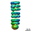
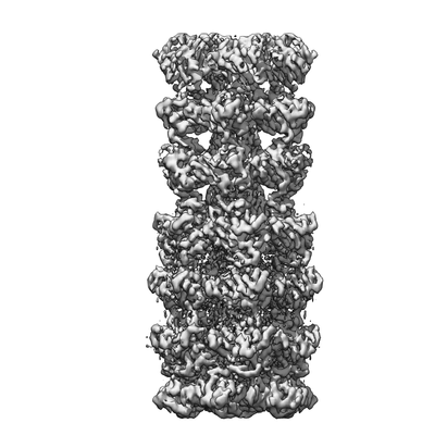
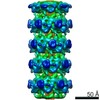
| Title | Lipophilic Envelope-spanning Tunnel B (LetB), Map 2 | ||||||||||||||||||
 Map data Map data | Lipophilic Envelope-spanning Tunnel B (LetB), Map 2 | ||||||||||||||||||
 Sample Sample |
| ||||||||||||||||||
 Keywords Keywords | lipid transport / bacterial cell envelope / MCE / YebT | ||||||||||||||||||
| Function / homology | : / Mce/MlaD / MlaD protein / intermembrane lipid transfer / membrane organization / outer membrane-bounded periplasmic space / identical protein binding / plasma membrane / Lipophilic envelope-spanning tunnel protein B Function and homology information Function and homology information | ||||||||||||||||||
| Biological species |  | ||||||||||||||||||
| Method | single particle reconstruction / cryo EM / Resolution: 3.49 Å | ||||||||||||||||||
 Authors Authors | Isom GL / Coudray N | ||||||||||||||||||
| Funding support |  United States, 5 items United States, 5 items
| ||||||||||||||||||
 Citation Citation | Journal: Cell / Year: 2020 Title: LetB Structure Reveals a Tunnel for Lipid Transport across the Bacterial Envelope. Authors: Georgia L Isom / Nicolas Coudray / Mark R MacRae / Collin T McManus / Damian C Ekiert / Gira Bhabha / Abstract: Gram-negative bacteria are surrounded by an outer membrane composed of phospholipids and lipopolysaccharide, which acts as a barrier and contributes to antibiotic resistance. The systems that mediate ...Gram-negative bacteria are surrounded by an outer membrane composed of phospholipids and lipopolysaccharide, which acts as a barrier and contributes to antibiotic resistance. The systems that mediate phospholipid trafficking across the periplasm, such as MCE (Mammalian Cell Entry) transporters, have not been well characterized. Our ~3.5 Å cryo-EM structure of the E. coli MCE protein LetB reveals an ~0.6 megadalton complex that consists of seven stacked rings, with a central hydrophobic tunnel sufficiently long to span the periplasm. Lipids bind inside the tunnel, suggesting that it functions as a pathway for lipid transport. Cryo-EM structures in the open and closed states reveal a dynamic tunnel lining, with implications for gating or substrate translocation. Our results support a model in which LetB establishes a physical link between the two membranes and creates a hydrophobic pathway for the translocation of lipids across the periplasm. | ||||||||||||||||||
| History |
|
- Structure visualization
| Movie |
Movie viewer |
|---|---|
| Structure viewer | EM map: SurfViewMolmilJmol/JSmol |
| Supplemental images |
- Downloads & links
-EMDB archive
| Map data | emd_20994.map.gz | 50.8 MB | EMDB map data format | |
|---|---|---|---|---|
| Header (meta data) | emd-20994-v30.xmlemd-20994.xml | 13.3 KB 13.3 KB | Display Display | EMDB header |
| Images |  emd_20994.png emd_20994.png | 50.6 KB | ||
| Filedesc metadata | emd-20994.cif.gz | 5.9 KB | ||
| Archive directory |  http://ftp.pdbj.org/pub/emdb/structures/EMD-20994ftp://ftp.pdbj.org/pub/emdb/structures/EMD-20994 http://ftp.pdbj.org/pub/emdb/structures/EMD-20994ftp://ftp.pdbj.org/pub/emdb/structures/EMD-20994 | HTTPS FTP |
-Related structure data
| Related structure data |  6v0dMC  6v0cC  6v0eC  6v0fC  6v0gC  6v0hC  6v0iC  6v0jC  6vciC M: atomic model generated by this map C: citing same article ( |
|---|---|
| Similar structure data | |
| EM raw data | EMPIAR-10350 (Title: Single particle cryo-EM dataset for LetB from E.coli / Data size: 928.6 Data #1: Aligned, dose-weighted and non-dose-weighted micrographs [micrographs - single frame] Data #2: Particle stacks for Model 1 (EMD-20993) [picked particles - single frame - processed] Data #3: Particle stacks for Model 2 (EMD-20994) [picked particles - single frame - processed] Data #4: Particle stacks for Model 3 (EMD-20995) [picked particles - single frame - processed] Data #5: Particle stacks for Model 4 (EMD-20996) [picked particles - single frame - processed] Data #6: Particle stacks for Model 5 (EMD-20997) [picked particles - single frame - processed] Data #7: Particle stacks for Model 6 (EMD-20998) [picked particles - single frame - processed] Data #8: Particle stacks for Model 7 (EMD-20999) [picked particles - single frame - processed] Data #9: Particle stacks for Model 8 (EMD-21000) [picked particles - single frame - processed]) |


-Links
| EMDB pages | EMDB (EBI/PDBe) / EMDataResource |
|---|
-Map
| File | Download / File: emd_20994.map.gz / Format: CCP4 / Size: 83.7 MB / Type: IMAGE STORED AS FLOATING POINT NUMBER (4 BYTES) | ||||||||||||||||||||||||||||||||||||||||||||||||||||||||||||
|---|---|---|---|---|---|---|---|---|---|---|---|---|---|---|---|---|---|---|---|---|---|---|---|---|---|---|---|---|---|---|---|---|---|---|---|---|---|---|---|---|---|---|---|---|---|---|---|---|---|---|---|---|---|---|---|---|---|---|---|---|---|
| Annotation | Lipophilic Envelope-spanning Tunnel B (LetB), Map 2 | ||||||||||||||||||||||||||||||||||||||||||||||||||||||||||||
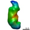
| Projections & slices | Image control
Images are generated by Spider. | ||||||||||||||||||||||||||||||||||||||||||||||||||||||||||||
| Voxel size | X=Y=Z: 1.31 Å | ||||||||||||||||||||||||||||||||||||||||||||||||||||||||||||
| Density |
| ||||||||||||||||||||||||||||||||||||||||||||||||||||||||||||
| Symmetry | Space group: 1 | ||||||||||||||||||||||||||||||||||||||||||||||||||||||||||||
| Details | EMDB XML:
CCP4 map header:
| ||||||||||||||||||||||||||||||||||||||||||||||||||||||||||||
 Z (Sec.)
 Y (Row.)
 X (Col.)


-Supplemental data
- Sample components
-Entire : Lipophilic Envelope-spanning Tunnel B (LetB), Map 2, Model 2
| Entire | Name: Lipophilic Envelope-spanning Tunnel B (LetB), Map 2, Model 2 |
|---|---|
| Components |
|
-Supramolecule #1: Lipophilic Envelope-spanning Tunnel B (LetB), Map 2, Model 2
| Supramolecule | Name: Lipophilic Envelope-spanning Tunnel B (LetB), Map 2, Model 2 type: complex / ID: 1 / Parent: 0 / Macromolecule list: all / Details: Cryo-EM reconstruction of full map, class 1.3 |
|---|---|
| Source (natural) | Organism: |
| Molecular weight | Theoretical: 560 KDa |
-Macromolecule #1: Intermembrane transport protein YebT
| Macromolecule | Name: Intermembrane transport protein YebT / type: protein_or_peptide / ID: 1 / Number of copies: 6 / Enantiomer: LEVO |
|---|---|
| Source (natural) | Organism: |
| Molecular weight | Theoretical: 89.744031 KDa |
| Recombinant expression | Organism: |
| Sequence | String: GNTVTIDFMS ADGIVPGRTP VRYQGVEVGT VQDISLSDDL RKIEVKVSIK SDMKDALREE TQFWLVTPKA SLAGVSGLDA LVGGNYIGM MPGKGKEQDH FVALDTQPKY RLDNGDLMIH LQAPDLGSLN SGSLVYFRKI PVGKVYDYAI NPNKQGVVID V LIERRFTD ...String: GNTVTIDFMS ADGIVPGRTP VRYQGVEVGT VQDISLSDDL RKIEVKVSIK SDMKDALREE TQFWLVTPKA SLAGVSGLDA LVGGNYIGM MPGKGKEQDH FVALDTQPKY RLDNGDLMIH LQAPDLGSLN SGSLVYFRKI PVGKVYDYAI NPNKQGVVID V LIERRFTD LVKKGSRFWN VSGVDANVSI SGAKVKLESL AALVNGAIAF DSPEESKPAE AEDTFGLYED LAHSQRGVII KL ELPSGAG LTADSTPLMY QGLEVGQLTK LDLNPGGKVT GEMTVDPSVV TLLRENTRIE LRNPKLSLSD ANLSALLTGK TFE LVPGDG EPRKEFVVVP GEKALLHEPD VLTLTLTAPE SYGIDAGQPL ILHGVQVGQV IDRKLTSKGV TFTVAIEPQH RELV KGDSK FVVNSRVDVK VGLDGVEFLG ASASEWINGG IRILPGDKGE MKASYPLYAN LEKALENSLS DLPTTTVSLS AETLP DVQA GSVVLYRKFE VGEVITVRPR ANAFDIDLHI KPEYRNLLTS NSVFWAEGGA KVQLNGSGLT VQASPLSRAL KGAISF DNL SGASASQRKG DKRILYASET AARAVGGQIT LHAFDAGKLA VGMPIRYLGI DIGQIQTLDL ITARNEVQAK AVLYPEY VQ TFARGGTRFS VVTPQISAAG VEHLDTILQP YINVEPGRGN PRRDFELQEA TITDSRYLDG LSIIVEAPEA GSLGIGTP V LFRGLEVGTV TGMTLGTLSD RVMIAMRISK RYQHLVRNNS VFWLASGYSL DFGLTGGVVK TGTFNQFIRG GIAFATPPG TPLAPKAQEG KHFLLQESEP KEWREWGTAL PK UniProtKB: Lipophilic envelope-spanning tunnel protein B |
-Experimental details
-Structure determination
| Method | cryo EM |
|---|---|
 Processing Processing | single particle reconstruction |
| Aggregation state | particle |
-Sample preparation
| Concentration | 0.5 mg/mL |
|---|---|
| Buffer | pH: 8 |
| Vitrification | Cryogen name: ETHANE / Instrument: FEI VITROBOT MARK III |
- Electron microscopy
| Microscope | FEI TITAN KRIOS |
|---|---|
| Image recording | Film or detector model: GATAN K2 SUMMIT (4k x 4k) / Digitization - Frames/image: 1-50 / Number grids imaged: 1 / Number real images: 4906 / Average electron dose: 80.0 e/Å2 |
| Electron beam | Acceleration voltage: 300 kV / Electron source:  FIELD EMISSION GUN FIELD EMISSION GUN |
| Electron optics | Illumination mode: FLOOD BEAM / Imaging mode: BRIGHT FIELD / Cs: 2.7 mm |
| Sample stage | Specimen holder model: FEI TITAN KRIOS AUTOGRID HOLDER |
| Experimental equipment |  Model: Titan Krios / Image courtesy: FEI Company |