Movie
Movie Controller


+ Open data

- Basic information
| Entry | Database: PDB / ID: 6v0d | ||||||||||||||||||
|---|---|---|---|---|---|---|---|---|---|---|---|---|---|---|---|---|---|---|---|
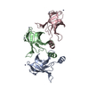
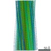
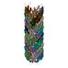
| Title | Lipophilic Envelope-spanning Tunnel B (LetB), Model 2 | ||||||||||||||||||
 Components Components | Intermembrane transport protein YebT | ||||||||||||||||||
 Keywords Keywords | LIPID TRANSPORT / bacterial cell envelope / MCE / YebT | ||||||||||||||||||
| Function / homology | : / Mce/MlaD / MlaD protein / intermembrane lipid transfer / membrane organization / outer membrane-bounded periplasmic space / identical protein binding / plasma membrane / Lipophilic envelope-spanning tunnel protein B Function and homology information Function and homology information | ||||||||||||||||||
| Biological species |  | ||||||||||||||||||
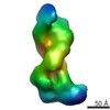
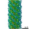
| Method | ELECTRON MICROSCOPY / single particle reconstruction / cryo EM / Resolution: 3.49 Å | ||||||||||||||||||
 Authors Authors | Isom, G.L. / Coudray, N. / MacRae, M.R. / McManus, C.T. / Ekiert, D.C. / Bhabha, G. | ||||||||||||||||||
| Funding support |  United States, 5items United States, 5items
| ||||||||||||||||||
 Citation Citation | Journal: Cell / Year: 2020 Title: LetB Structure Reveals a Tunnel for Lipid Transport across the Bacterial Envelope. Authors: Georgia L Isom / Nicolas Coudray / Mark R MacRae / Collin T McManus / Damian C Ekiert / Gira Bhabha / Abstract: Gram-negative bacteria are surrounded by an outer membrane composed of phospholipids and lipopolysaccharide, which acts as a barrier and contributes to antibiotic resistance. The systems that mediate ...Gram-negative bacteria are surrounded by an outer membrane composed of phospholipids and lipopolysaccharide, which acts as a barrier and contributes to antibiotic resistance. The systems that mediate phospholipid trafficking across the periplasm, such as MCE (Mammalian Cell Entry) transporters, have not been well characterized. Our ~3.5 Å cryo-EM structure of the E. coli MCE protein LetB reveals an ~0.6 megadalton complex that consists of seven stacked rings, with a central hydrophobic tunnel sufficiently long to span the periplasm. Lipids bind inside the tunnel, suggesting that it functions as a pathway for lipid transport. Cryo-EM structures in the open and closed states reveal a dynamic tunnel lining, with implications for gating or substrate translocation. Our results support a model in which LetB establishes a physical link between the two membranes and creates a hydrophobic pathway for the translocation of lipids across the periplasm. | ||||||||||||||||||
| History |
|
- Structure visualization
| Movie |
Movie viewer |
|---|---|
| Structure viewer | Molecule: MolmilJmol/JSmol |
- Downloads & links
-Download
| PDBx/mmCIF format | 6v0d.cif.gz | 802.2 KB | Display | PDBx/mmCIF format |
|---|---|---|---|---|
| PDB format | pdb6v0d.ent.gz | 676.4 KB | Display | PDB format |
| PDBx/mmJSON format | 6v0d.json.gz | Tree view | PDBx/mmJSON format | |
| Others |  Other downloads Other downloads |
-Validation report
| Arichive directory | https://data.pdbj.org/pub/pdb/validation_reports/v0/6v0dftp://data.pdbj.org/pub/pdb/validation_reports/v0/6v0d | HTTPS FTP |
|---|
-Related structure data
| Related structure data | 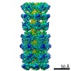 20994MC  6v0cC  6v0eC  6v0fC  6v0gC  6v0hC  6v0iC  6v0jC  6vciC M: map data used to model this data C: citing same article ( |
|---|---|
| Similar structure data | |
| EM raw data | EMPIAR-10350 (Title: Single particle cryo-EM dataset for LetB from E.coli / Data size: 928.6 Data #1: Aligned, dose-weighted and non-dose-weighted micrographs [micrographs - single frame] Data #2: Particle stacks for Model 1 (EMD-20993) [picked particles - single frame - processed] Data #3: Particle stacks for Model 2 (EMD-20994) [picked particles - single frame - processed] Data #4: Particle stacks for Model 3 (EMD-20995) [picked particles - single frame - processed] Data #5: Particle stacks for Model 4 (EMD-20996) [picked particles - single frame - processed] Data #6: Particle stacks for Model 5 (EMD-20997) [picked particles - single frame - processed] Data #7: Particle stacks for Model 6 (EMD-20998) [picked particles - single frame - processed] Data #8: Particle stacks for Model 7 (EMD-20999) [picked particles - single frame - processed] Data #9: Particle stacks for Model 8 (EMD-21000) [picked particles - single frame - processed]) |


-Links
 PDBj
PDBj- Assembly
| Deposited unit | 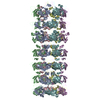
|
|---|---|
| 1 |
|
-Components
| #1: Protein | Mass: 89744.031 Da / Num. of mol.: 6 Source method: isolated from a genetically manipulated source Source: (gene. exp.) Strain: K12 / Gene: yebT, b1834, JW1823 / Production host: |
|---|
-Experimental details
-Experiment
| Experiment | Method: ELECTRON MICROSCOPY |
|---|---|
| EM experiment | Aggregation state: PARTICLE / 3D reconstruction method: single particle reconstruction |
- Sample preparation
| Component | Name: Lipophilic Envelope-spanning Tunnel B (LetB), Map 2, Model 2 Type: COMPLEX / Details: Cryo-EM reconstruction of full map, class 1.3 / Entity ID: all / Source: NATURAL |
|---|---|
| Molecular weight | Value: 0.56 MDa / Experimental value: NO |
| Source (natural) | Organism: |
| Buffer solution | pH: 8 |
| Specimen | Conc.: 0.5 mg/ml / Embedding applied: NO / Shadowing applied: NO / Staining applied: NO / Vitrification applied: YES |
| Vitrification | Instrument: FEI VITROBOT MARK III / Cryogen name: ETHANE |
- Electron microscopy imaging
| Experimental equipment |  Model: Titan Krios / Image courtesy: FEI Company |
|---|---|
| Microscopy | Model: FEI TITAN KRIOS |
| Electron gun | Electron source:  FIELD EMISSION GUN / Accelerating voltage: 300 kV / Illumination mode: FLOOD BEAM FIELD EMISSION GUN / Accelerating voltage: 300 kV / Illumination mode: FLOOD BEAM |
| Electron lens | Mode: BRIGHT FIELD / Cs: 2.7 mm |
| Specimen holder | Specimen holder model: FEI TITAN KRIOS AUTOGRID HOLDER |
| Image recording | Electron dose: 80 e/Å2 / Film or detector model: GATAN K2 SUMMIT (4k x 4k) / Num. of grids imaged: 1 / Num. of real images: 4906 |
| Image scans | Movie frames/image: 50 / Used frames/image: 1-50 |
- Processing
| EM software |
| ||||||||||||||||||||||||||||||||||||||||
|---|---|---|---|---|---|---|---|---|---|---|---|---|---|---|---|---|---|---|---|---|---|---|---|---|---|---|---|---|---|---|---|---|---|---|---|---|---|---|---|---|---|
| CTF correction | Type: PHASE FLIPPING AND AMPLITUDE CORRECTION | ||||||||||||||||||||||||||||||||||||||||
| Particle selection | Num. of particles selected: 731231 | ||||||||||||||||||||||||||||||||||||||||
| Symmetry | Point symmetry: C6 (6 fold cyclic) | ||||||||||||||||||||||||||||||||||||||||
| 3D reconstruction | Resolution: 3.49 Å / Resolution method: FSC 0.143 CUT-OFF / Num. of particles: 57157 / Symmetry type: POINT | ||||||||||||||||||||||||||||||||||||||||
| Atomic model building | PDB-ID: 5UW2 Pdb chain-ID: A / Accession code: 5UW2 / Source name: PDB / Type: experimental model |