 Movie
Movie Controller


+ Open data

- Basic information
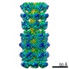
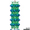
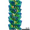
| Entry | Database: PDB / ID: 5leg | ||||||||||||
|---|---|---|---|---|---|---|---|---|---|---|---|---|---|
| Title | Structure of the bacterial sex F pilus (pED208) | ||||||||||||
 Components Components | Pilin | ||||||||||||
 Keywords Keywords | PROTEIN FIBRIL / F-pilus Conjugation Type IV secretion system Phospholipid | ||||||||||||
| Function / homology | TraA / TraA / extracellular region / plasma membrane / 1,2-DIPALMITOYL-PHOSPHATIDYL-GLYCEROLE / Pilin Function and homology information Function and homology information | ||||||||||||
| Biological species |  Salmonella enterica subsp. enterica serovar Typhi (bacteria) Salmonella enterica subsp. enterica serovar Typhi (bacteria) | ||||||||||||
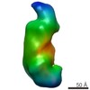
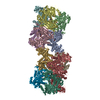
| Method | ELECTRON MICROSCOPY / helical reconstruction / cryo EM / Resolution: 3.6 Å | ||||||||||||
 Authors Authors | Costa, T.R.D. / Ilangovan, I. / Ukleja, M. / Redzej, A. / Santini, J.M. / Smith, T.K. / Egelman, E.H. / Waksman, G. | ||||||||||||
| Funding support |  United Kingdom, United Kingdom,  United States, 3items United States, 3items
| ||||||||||||
 Citation Citation | Journal: Cell / Year: 2016 Title: Structure of the Bacterial Sex F Pilus Reveals an Assembly of a Stoichiometric Protein-Phospholipid Complex. Authors: Tiago R D Costa / Aravindan Ilangovan / Marta Ukleja / Adam Redzej / Joanne M Santini / Terry K Smith / Edward H Egelman / Gabriel Waksman / Abstract: Conjugative pili are widespread bacterial appendages that play important roles in horizontal gene transfer, in spread of antibiotic resistance genes, and as sites of phage attachment. Among ...Conjugative pili are widespread bacterial appendages that play important roles in horizontal gene transfer, in spread of antibiotic resistance genes, and as sites of phage attachment. Among conjugative pili, the F "sex" pilus encoded by the F plasmid is the best functionally characterized, and it is also historically the most important, as the discovery of F-plasmid-mediated conjugation ushered in the era of molecular biology and genetics. Yet, its structure is unknown. Here, we present atomic models of two F family pili, the F and pED208 pili, generated from cryoelectron microscopy reconstructions at 5.0 and 3.6 Å resolution, respectively. These structures reveal that conjugative pili are assemblies of stoichiometric protein-phospholipid units. We further demonstrate that each pilus type binds preferentially to particular phospholipids. These structures provide the molecular basis for F pilus assembly and also shed light on the remarkable properties of conjugative pili in bacterial secretion and phage infection. | ||||||||||||
| History |
|
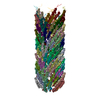
- Structure visualization
| Movie |
Movie viewer |
|---|---|
| Structure viewer | Molecule: MolmilJmol/JSmol |
- Downloads & links
-Download
| PDBx/mmCIF format | 5leg.cif.gz | 863.6 KB | Display | PDBx/mmCIF format |
|---|---|---|---|---|
| PDB format | pdb5leg.ent.gz | Display | PDB format | |
| PDBx/mmJSON format | 5leg.json.gz | Tree view | PDBx/mmJSON format | |
| Others |  Other downloads Other downloads |
-Validation report
| Arichive directory | https://data.pdbj.org/pub/pdb/validation_reports/le/5legftp://data.pdbj.org/pub/pdb/validation_reports/le/5leg | HTTPS FTP |
|---|
-Related structure data
| Related structure data | 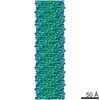 4042MC 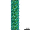 4044C 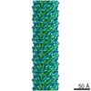 4046C 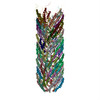 5lerC 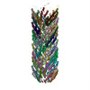 5lfbC M: map data used to model this data C: citing same article ( |
|---|---|
| Similar structure data |


-Links
 PDBj
PDBj


- Assembly
| Deposited unit | 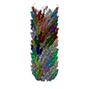
|
|---|---|
| 1 |
|
-Components
| #1: Protein | Mass: 6750.250 Da / Num. of mol.: 80 / Fragment: UNP Residues 57-119 / Source method: isolated from a natural source Source: (natural) Salmonella enterica subsp. enterica serovar Typhi (bacteria)References: UniProt: P12060 #2: Chemical | ChemComp-LHG /   Mass: 722.970 Da / Num. of mol.: 75 / Source method: obtained synthetically / Formula: C38H75O10P / Comment: phospholipid*YM Mass: 722.970 Da / Num. of mol.: 75 / Source method: obtained synthetically / Formula: C38H75O10P / Comment: phospholipid*YM |
|---|
-Experimental details
-Experiment
| Experiment | Method: ELECTRON MICROSCOPY |
|---|---|
| EM experiment | Aggregation state: HELICAL ARRAY / 3D reconstruction method: helical reconstruction |
- Sample preparation
| Component | Name: F-pilus (pED208)filament made by an assembly of a stoichiometric protein-phospholipid complex Type: ORGANELLE OR CELLULAR COMPONENT / Entity ID: #1 / Source: NATURAL |
|---|---|
| Molecular weight | Value: 6.3 kDa/nm / Experimental value: NO |
| Source (natural) | Organism: |
| Buffer solution | pH: 7.4 |
| Specimen | Embedding applied: NO / Shadowing applied: NO / Staining applied: NO / Vitrification applied: YES |
| Specimen support | Grid material: COPPER / Grid mesh size: 400 divisions/in. / Grid type: Lacey |
| Vitrification | Cryogen name: ETHANE |
- Electron microscopy imaging
| Experimental equipment |  Model: Tecnai Polara / Image courtesy: FEI Company |
|---|---|
| Microscopy | Model: FEI POLARA 300 |
| Electron gun | Electron source:  FIELD EMISSION GUN / Accelerating voltage: 300 kV / Illumination mode: FLOOD BEAM FIELD EMISSION GUN / Accelerating voltage: 300 kV / Illumination mode: FLOOD BEAM |
| Electron lens | Mode: BRIGHT FIELD |
| Image recording | Electron dose: 1.67 e/Å2 / Film or detector model: GATAN K2 SUMMIT (4k x 4k) |
- Processing
| EM software |
| |||||||||||||||||||||||||||||||||||||||||||||||||||||||
|---|---|---|---|---|---|---|---|---|---|---|---|---|---|---|---|---|---|---|---|---|---|---|---|---|---|---|---|---|---|---|---|---|---|---|---|---|---|---|---|---|---|---|---|---|---|---|---|---|---|---|---|---|---|---|---|---|
| CTF correction | Type: PHASE FLIPPING AND AMPLITUDE CORRECTION | |||||||||||||||||||||||||||||||||||||||||||||||||||||||
| Helical symmerty | Angular rotation/subunit: 28.2 ° / Axial rise/subunit: 12.1 Å / Axial symmetry: C5 | |||||||||||||||||||||||||||||||||||||||||||||||||||||||
| 3D reconstruction | Resolution: 3.6 Å / Resolution method: OTHER / Num. of particles: 43952 / Symmetry type: HELICAL | |||||||||||||||||||||||||||||||||||||||||||||||||||||||
| Atomic model building | Protocol: AB INITIO MODEL |
