National Institutes of Health/National Institute of General Medical Sciences (NIH/NIGMS)
GM122510
United States
Swiss National Science Foundation
310030_144243
Switzerland
Citation
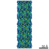
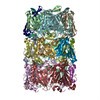
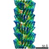
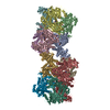
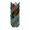
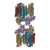
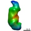
Journal: Nat Struct Mol Biol / Year: 2015 Title: Atomic structures of a bactericidal contractile nanotube in its pre- and postcontraction states. Authors: Peng Ge / Dean Scholl / Petr G Leiman / Xuekui Yu / Jeff F Miller / Z Hong Zhou / Abstract: R-type pyocins are representatives of contractile ejection systems, a class of biological nanomachines that includes, among others, the bacterial type VI secretion system (T6SS) and contractile ...R-type pyocins are representatives of contractile ejection systems, a class of biological nanomachines that includes, among others, the bacterial type VI secretion system (T6SS) and contractile bacteriophage tails. We report atomic models of the Pseudomonas aeruginosa precontraction pyocin sheath and tube, and the postcontraction sheath, obtained by cryo-EM at 3.5-Å and 3.9-Å resolutions, respectively. The central channel of the tube is negatively charged, in contrast to the neutral and positive counterparts in T6SSs and phage tails. The sheath is interwoven by long N- and C-terminal extension arms emanating from each subunit, which create an extensive two-dimensional mesh that has the same connectivity in the extended and contracted state of the sheath. We propose that the contraction process draws energy from electrostatic and shape complementarities to insert the inner tube through bacterial cell membranes to eventually kill the bacteria.
THIS ENTRY 5W5E REFLECTS AN ALTERNATIVE MODELING OF THE ORIGINAL DATA IN EMD-6270, DETERMINED BY P. ...THIS ENTRY 5W5E REFLECTS AN ALTERNATIVE MODELING OF THE ORIGINAL DATA IN EMD-6270, DETERMINED BY P.GE, D.SCHOLL, P.G.LEIMAN, X.YU, J.F.MILLER, Z.H.ZHOU
A: FIIR2 protein B: FIIR2 protein C: FIIR2 protein D: FIIR2 protein E: FIIR2 protein F: FIIR2 protein G: FIIR2 protein H: FIIR2 protein I: FIIR2 protein J: FIIR2 protein K: FIIR2 protein L: FIIR2 protein M: FIIR2 protein N: FIIR2 protein O: FIIR2 protein P: FIIR2 protein Q: FIIR2 protein R: FIIR2 protein S: FIIR2 protein T: FIIR2 protein U: FIIR2 protein V: FIIR2 protein W: FIIR2 protein X: FIIR2 protein Y: FIIR2 protein Z: FIIR2 protein a: FIIR2 protein b: FIIR2 protein c: FIIR2 protein d: FIIR2 protein
Helical symmetry: (Circular symmetry: 6 / Dyad axis: no / N subunits divisor: 1 / Num. of operations: 30 / Rise per n subunits: 38.4 Å / Rotation per n subunits: 18.3 °)
Details
A helical assembly can be generated by applying the helical parameters to a single subunit, e.g., chain A.
-
Components
#1: Protein
... FIIR2protein / pyocin tube
Mass: 18088.547 Da / Num. of mol.: 30 Source method: isolated from a genetically manipulated source Source: (gene. exp.) Pseudomonas aeruginosa (bacteria) / Gene: FIIR2 / Production host: Pseudomonas aeruginosa (bacteria) / References: UniProt: Q9S573
-
Experimental details
-
Experiment
Experiment
Method: ELECTRON MICROSCOPY
EM experiment
Aggregation state: FILAMENT / 3D reconstruction method: helical reconstruction
-
Sample preparation
Component
Name: pyocin tube of Pseudomonas aeruginosa / Type: COMPLEX / Entity ID: all / Source: NATURAL
Source (natural)
Organism: Pseudomonas aeruginosa (bacteria)
Specimen
Embedding applied: NO / Shadowing applied: NO / Staining applied: NO / Vitrification applied: YES
Vitrification
Cryogen name: ETHANE
-
Electron microscopy imaging
Experimental equipment
Model: Titan Krios / Image courtesy: FEI Company
Microscopy
Model: FEI TITAN KRIOS
Electron gun
Electron source: FIELD EMISSION GUN / Accelerating voltage: 300 kV / Illumination mode: FLOOD BEAM
In the structure databanks used in Yorodumi, some data are registered as the other names, "COVID-19 virus" and "2019-nCoV". Here are the details of the virus and the list of structure data.
Jan 31, 2019. EMDB accession codes are about to change! (news from PDBe EMDB page)
EMDB accession codes are about to change! (news from PDBe EMDB page)
The allocation of 4 digits for EMDB accession codes will soon come to an end. Whilst these codes will remain in use, new EMDB accession codes will include an additional digit and will expand incrementally as the available range of codes is exhausted. The current 4-digit format prefixed with “EMD-” (i.e. EMD-XXXX) will advance to a 5-digit format (i.e. EMD-XXXXX), and so on. It is currently estimated that the 4-digit codes will be depleted around Spring 2019, at which point the 5-digit format will come into force.
The EM Navigator/Yorodumi systems omit the EMD- prefix.
Related info.:Q: What is EMD? / ID/Accession-code notation in Yorodumi/EM Navigator
Yorodumi is a browser for structure data from EMDB, PDB, SASBDB, etc.
This page is also the successor to EM Navigator detail page, and also detail information page/front-end page for Omokage search.
The word "yorodu" (or yorozu) is an old Japanese word meaning "ten thousand". "mi" (miru) is to see.
Related info.:EMDB / PDB / SASBDB / Comparison of 3 databanks / Yorodumi Search / Aug 31, 2016. New EM Navigator & Yorodumi / Yorodumi Papers / Jmol/JSmol / Function and homology information / Changes in new EM Navigator and Yorodumi
 Movie
Movie Controller


 Open data
 Basic information
 Components
 Keywords
 Function and homology information
 Pseudomonas aeruginosa (bacteria)
 Authors
 United States,
  Switzerland, 2items
  Citation
 Structure visualization
 Downloads & links
 Other downloads
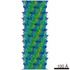


 PDBj
PDBj Assembly
 Sample preparation
 Electron microscopy imaging
 FIELD EMISSION GUN / Accelerating voltage: 300 kV / Illumination mode: FLOOD BEAM
 Processing