 Movie
Movie Controller


+ Open data

- Basic information
| Entry | Database: EMDB / ID: EMD-8767 | |||||||||
|---|---|---|---|---|---|---|---|---|---|---|
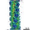
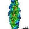
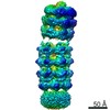
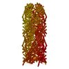
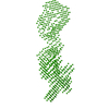
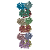
| Title | Cryo-EM structure of the T4 tail tube | |||||||||
 Map data Map data | Enterobacteria Phage T4 sensu lato tail tube protein gp19 filament | |||||||||
 Sample Sample |
| |||||||||
 Keywords Keywords | T4 tail tube / dose limit / helical reconstruction / VIRAL PROTEIN | |||||||||
| Function / homology | symbiont genome ejection through host cell envelope, contractile tail mechanism / Bacteriophage T4, Gp19, tail tube / T4-like virus tail tube protein gp19 / virus tail, tube / structural molecule activity / Tail tube protein gp19 Function and homology information Function and homology information | |||||||||
| Biological species |  Enterobacteria phage T4 sensu lato (virus) Enterobacteria phage T4 sensu lato (virus) | |||||||||
| Method | helical reconstruction / cryo EM / Resolution: 3.4 Å | |||||||||
 Authors Authors | Zheng W / Wang F | |||||||||
| Funding support |  United States, United States,  Switzerland, 2 items Switzerland, 2 items
| |||||||||
 Citation Citation | Journal: Structure / Year: 2017 Title: Refined Cryo-EM Structure of the T4 Tail Tube: Exploring the Lowest Dose Limit. Authors: Weili Zheng / Fengbin Wang / Nicholas M I Taylor / Ricardo C Guerrero-Ferreira / Petr G Leiman / Edward H Egelman / Abstract: The bacteriophage T4 contractile tail (containing a tube and sheath) was the first biological assembly reconstructed in three dimensions by electron microscopy at a resolution of ∼35 Å in 1968. A ...The bacteriophage T4 contractile tail (containing a tube and sheath) was the first biological assembly reconstructed in three dimensions by electron microscopy at a resolution of ∼35 Å in 1968. A single-particle reconstruction of the T4 baseplate was able to generate a 4.1 Å resolution map for the first two rings of the tube using the overall baseplate for alignment. We have now reconstructed the T4 tail tube at a resolution of 3.4 Å, more than a 1,000-fold increase in information content for the tube from 1968. We have used legacy software (Spider) to show that we can do better than the typical 2/3 Nyquist frequency. A reasonable map can be generated with only 1.5 electrons/Å using the higher dose images for alignment, but increasing the dose results in a better map, consistent with other reports that electron dose does not represent the main limitation on resolution in cryo-electron microscopy. | |||||||||
| History |
|
- Structure visualization
| Movie |
Movie viewer |
|---|---|
| Structure viewer | EM map: SurfViewMolmilJmol/JSmol |
| Supplemental images |

- Downloads & links
-EMDB archive
| Map data | emd_8767.map.gz | 3.3 MB | EMDB map data format | |
|---|---|---|---|---|
| Header (meta data) | emd-8767-v30.xmlemd-8767.xml | 10.9 KB 10.9 KB | Display Display | EMDB header |
| Images | 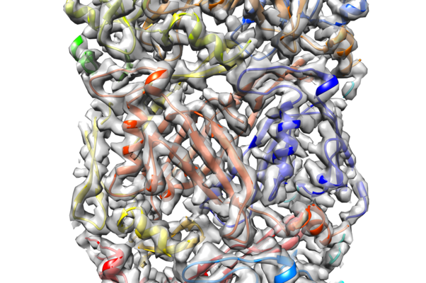 emd_8767.png emd_8767.png | 318 KB | ||
| Filedesc metadata | emd-8767.cif.gz | 5.2 KB | ||
| Archive directory |  http://ftp.pdbj.org/pub/emdb/structures/EMD-8767ftp://ftp.pdbj.org/pub/emdb/structures/EMD-8767 http://ftp.pdbj.org/pub/emdb/structures/EMD-8767ftp://ftp.pdbj.org/pub/emdb/structures/EMD-8767 | HTTPS FTP |
-Related structure data
| Related structure data |  5w5fMC  5w5eC M: atomic model generated by this map C: citing same article ( |
|---|---|
| Similar structure data |
-Links
| EMDB pages | EMDB (EBI/PDBe) / EMDataResource |
|---|
-Map
| File | Download / File: emd_8767.map.gz / Format: CCP4 / Size: 7.6 MB / Type: IMAGE STORED AS FLOATING POINT NUMBER (4 BYTES) | ||||||||||||||||||||||||||||||||||||||||||||||||||||||||||||
|---|---|---|---|---|---|---|---|---|---|---|---|---|---|---|---|---|---|---|---|---|---|---|---|---|---|---|---|---|---|---|---|---|---|---|---|---|---|---|---|---|---|---|---|---|---|---|---|---|---|---|---|---|---|---|---|---|---|---|---|---|---|
| Annotation | Enterobacteria Phage T4 sensu lato tail tube protein gp19 filament | ||||||||||||||||||||||||||||||||||||||||||||||||||||||||||||
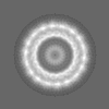
| Projections & slices | Image control
Images are generated by Spider. generated in cubic-lattice coordinate | ||||||||||||||||||||||||||||||||||||||||||||||||||||||||||||
| Voxel size | X=Y=Z: 1.32 Å | ||||||||||||||||||||||||||||||||||||||||||||||||||||||||||||
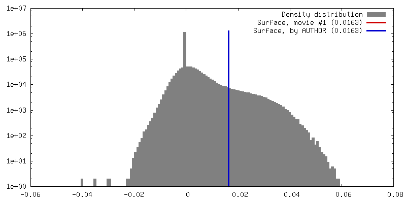
| Density |
| ||||||||||||||||||||||||||||||||||||||||||||||||||||||||||||
| Symmetry | Space group: 1 | ||||||||||||||||||||||||||||||||||||||||||||||||||||||||||||
| Details | EMDB XML:
CCP4 map header:
| ||||||||||||||||||||||||||||||||||||||||||||||||||||||||||||
 Z (Sec.)
 Y (Row.)
 X (Col.)


-Supplemental data
- Sample components
-Entire : Enterobacteria Phage T4 sensu lato tail tube protein gp19 filament
| Entire | Name: Enterobacteria Phage T4 sensu lato tail tube protein gp19 filament |
|---|---|
| Components |
|
-Supramolecule #1: Enterobacteria Phage T4 sensu lato tail tube protein gp19 filament
| Supramolecule | Name: Enterobacteria Phage T4 sensu lato tail tube protein gp19 filament type: complex / ID: 1 / Parent: 0 / Macromolecule list: all |
|---|---|
| Source (natural) | Organism: Enterobacteria phage T4 sensu lato (virus) |
-Macromolecule #1: Tail tube protein gp19
| Macromolecule | Name: Tail tube protein gp19 / type: protein_or_peptide / ID: 1 / Number of copies: 18 / Enantiomer: LEVO |
|---|---|
| Source (natural) | Organism: Enterobacteria phage T4 sensu lato (virus) |
| Molecular weight | Theoretical: 18.479613 KDa |
| Sequence | String: MFVDDVTRAF ESGDFARPNL FQVEISYLGQ NFTFQCKATA LPAGIVEKIP VGFMNRKINV AGDRTFDDWT VTVMNDEAHD ARQKFVDWQ SIAAGQGNEI TGGKPAEYKK SAIVRQYARD AKTVTKEIEI KGLWPTNVGE LQLDWDSNNE IQTFEVTLAL D YWE UniProtKB: Tail tube protein gp19 |
-Experimental details
-Structure determination
| Method | cryo EM |
|---|---|
 Processing Processing | helical reconstruction |
| Aggregation state | filament |
-Sample preparation
| Concentration | 1 mg/mL |
|---|---|
| Buffer | pH: 8 |
| Grid | Model: Quantifoil / Material: COPPER / Mesh: 300 / Pretreatment - Type: GLOW DISCHARGE |
| Vitrification | Cryogen name: ETHANE / Instrument: FEI VITROBOT MARK IV |
- Electron microscopy
| Microscope | FEI TITAN KRIOS |
|---|---|
| Specialist optics | Energy filter - Name: Gatan image filter |
| Image recording | Film or detector model: GATAN K2 SUMMIT (4k x 4k) / Average electron dose: 40.0 e/Å2 |
| Electron beam | Acceleration voltage: 300 kV / Electron source:  FIELD EMISSION GUN FIELD EMISSION GUN |
| Electron optics | Illumination mode: FLOOD BEAM / Imaging mode: BRIGHT FIELD |
| Experimental equipment |  Model: Titan Krios / Image courtesy: FEI Company |
-Image processing
| Final reconstruction | Applied symmetry - Helical parameters - Δz: 40.2 Å Applied symmetry - Helical parameters - Δ&Phi: 18.2 ° Applied symmetry - Helical parameters - Axial symmetry: C6 (6 fold cyclic) Resolution.type: BY AUTHOR / Resolution: 3.4 Å / Resolution method: FSC 0.143 CUT-OFF / Software - Name: SPIDER / Number images used: 26320 |
|---|---|
| Segment selection | Number selected: 26320 / Software - Name: EMAN2 |
| Startup model | Type of model: NONE / Details: featureless cylinder |
| Final angle assignment | Type: NOT APPLICABLE / Software - Name: SPIDER |
-Atomic model buiding 1
| Refinement | Space: REAL |
|---|---|
| Output model | PDB-5w5f: |
