 Movie
Movie Controller


+ Open data

- Basic information
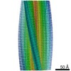
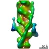
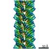
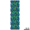
| Entry | Database: PDB / ID: 3j4s | ||||||
|---|---|---|---|---|---|---|---|
| Title | Helical Model of TubZ-Bt four-stranded filament | ||||||
 Components Components | FtsZ/tubulin-related protein | ||||||
 Keywords Keywords | STRUCTURAL PROTEIN / FtsZ-like / Tubulin-like / GTPase | ||||||
| Function / homology |  Function and homology information Function and homology informationplasmid partitioning / Hydrolases; Acting on acid anhydrides; Acting on GTP to facilitate cellular and subcellular movement / GTPase activity / GTP binding / metal ion binding / identical protein binding / cytoplasm Similarity search - Function | ||||||
| Biological species |  | ||||||
| Method | ELECTRON MICROSCOPY / helical reconstruction / cryo EM / Resolution: 6.8 Å | ||||||
 Authors Authors | Montabana, E.A. / Agard, D.A. | ||||||
 Citation Citation | Journal: Proc Natl Acad Sci U S A / Year: 2014 Title: Bacterial tubulin TubZ-Bt transitions between a two-stranded intermediate and a four-stranded filament upon GTP hydrolysis. Authors: Elizabeth A Montabana / David A Agard /  Abstract: Cytoskeletal filaments form diverse superstructures that are highly adapted for specific functions. The recently discovered TubZ subfamily of tubulins is involved in type III plasmid partitioning ...Cytoskeletal filaments form diverse superstructures that are highly adapted for specific functions. The recently discovered TubZ subfamily of tubulins is involved in type III plasmid partitioning systems, facilitating faithful segregation of low copy-number plasmids during bacterial cell division. One such protein, TubZ-Bt, is found on the large pBtoxis plasmid in Bacillus thuringiensis, and interacts via its extended C terminus with a DNA adaptor protein TubR. Here, we use cryo-electron microscopy to determine the structure of TubZ-Bt filaments and light scattering to explore their mechanism of polymerization. Surprisingly, we find that the helical filament architecture is remarkably sensitive to nucleotide state, changing from two-stranded to four-stranded depending on the ability of TubZ-Bt to hydrolyze GTP. We present pseudoatomic models of both the two- and four-protofilament forms based on cryo-electron microscopy reconstructions (10.8 Å and 6.9 Å, respectively) of filaments formed under different nucleotide states. These data lead to a model in which the two-stranded filament is a necessary intermediate along the pathway to formation of the four-stranded filament. Such nucleotide-directed structural polymorphism is to our knowledge an unprecedented mechanism for the formation of polar filaments. | ||||||
| History |
|
- Structure visualization
| Movie |
Movie viewer |
|---|---|
| Structure viewer | Molecule: MolmilJmol/JSmol |
- Downloads & links
-Download
| PDBx/mmCIF format | 3j4s.cif.gz | 86.4 KB | Display | PDBx/mmCIF format |
|---|---|---|---|---|
| PDB format | pdb3j4s.ent.gz | 62.2 KB | Display | PDB format |
| PDBx/mmJSON format | 3j4s.json.gz | Tree view | PDBx/mmJSON format | |
| Others |  Other downloads Other downloads |
-Validation report
| Arichive directory | https://data.pdbj.org/pub/pdb/validation_reports/j4/3j4sftp://data.pdbj.org/pub/pdb/validation_reports/j4/3j4s | HTTPS FTP |
|---|
-Related structure data
| Related structure data | 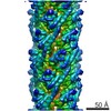 5762MC 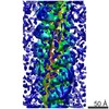 5763C  3j4tC M: map data used to model this data C: citing same article ( |
|---|---|
| Similar structure data |


-Links
 PDBj
PDBj


- Assembly
| Deposited unit | 
|
|---|---|
| 1 | x 24
|
| 2 |
|
| 3 | 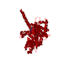
|
| Symmetry | Helical symmetry: (Circular symmetry: 4 / Dyad axis: no / N subunits divisor: 1 / Num. of operations: 24 / Rise per n subunits: 43.545 Å / Rotation per n subunits: 31.788 °) |
-Components
| #1: Protein | Mass: 54435.785 Da / Num. of mol.: 1 / Mutation: L2V Source method: isolated from a genetically manipulated source Source: (gene. exp.) |
|---|---|
| #2: Chemical | ChemComp-GDP /   Type: RNA linking / Mass: 443.201 Da / Num. of mol.: 1 / Source method: obtained synthetically / Formula: C10H15N5O11P2 / Comment: GDP, energy-carrying molecule*YM Type: RNA linking / Mass: 443.201 Da / Num. of mol.: 1 / Source method: obtained synthetically / Formula: C10H15N5O11P2 / Comment: GDP, energy-carrying molecule*YM |
-Experimental details
-Experiment
| Experiment | Method: ELECTRON MICROSCOPY |
|---|---|
| EM experiment | Aggregation state: FILAMENT / 3D reconstruction method: helical reconstruction |
- Sample preparation
| Component | Name: TubZ-Bt / Type: COMPLEX |
|---|---|
| Buffer solution | Name: HMK100 / pH: 7.7 Details: 100 mM potassium acetate, 5 mM magnesium acetate, 50 mM HEPES |
| Specimen | Embedding applied: NO / Shadowing applied: NO / Staining applied: NO / Vitrification applied: YES |
| Specimen support | Details: 400 mesh copper grid with holey carbon support, glow discharged |
| Vitrification | Instrument: FEI VITROBOT MARK III / Cryogen name: ETHANE / Humidity: 100 % Details: Add GTP to grow filaments for 6 minutes before application to grid. Blot 4.5 seconds before plunging into liquid ethane (FEI VITROBOT MARK III). Method: Blot 4.5 s before plunging |
- Electron microscopy imaging
| Experimental equipment |  Model: Tecnai F20 / Image courtesy: FEI Company |
|---|---|
| Microscopy | Model: FEI TECNAI F20 / Date: Apr 22, 2011 |
| Electron gun | Electron source:  FIELD EMISSION GUN / Accelerating voltage: 200 kV / Illumination mode: FLOOD BEAM FIELD EMISSION GUN / Accelerating voltage: 200 kV / Illumination mode: FLOOD BEAM |
| Electron lens | Mode: BRIGHT FIELD / Nominal magnification: 62000 X / Nominal defocus max: 3500 nm / Nominal defocus min: 800 nm / Cs: 2.2 mm |
| Specimen holder | Specimen holder model: OTHER / Specimen holder type: Oxford side-entry cryo stage |
| Image recording | Electron dose: 20 e/Å2 / Film or detector model: TVIPS TEMCAM-F816 (8k x 8k) / Details: 8K x 8K |
| Image scans | Num. digital images: 414 |
| Radiation | Protocol: SINGLE WAVELENGTH / Monochromatic (M) / Laue (L): M / Scattering type: x-ray |
| Radiation wavelength | Relative weight: 1 |
- Processing
| EM software |
| |||||||||||||||
|---|---|---|---|---|---|---|---|---|---|---|---|---|---|---|---|---|
| CTF correction | Details: Whole Micrograph | |||||||||||||||
| Helical symmerty | Angular rotation/subunit: 31.78843 ° / Axial rise/subunit: 43.54512 Å / Axial symmetry: C4 | |||||||||||||||
| 3D reconstruction | Method: Iterative Helical Real Space Refinement / Resolution: 6.8 Å / Resolution method: FSC 0.143 CUT-OFF / Actual pixel size: 0.94 Å Details: Final map has been low-pass filtered to 7 Angstrom and high-pass filtered to 30 Angstrom. A B-factor of -309 Angstrom was applied using the program bfactor. A cylindrical mask of radius ~80 ...Details: Final map has been low-pass filtered to 7 Angstrom and high-pass filtered to 30 Angstrom. A B-factor of -309 Angstrom was applied using the program bfactor. A cylindrical mask of radius ~80 Angstrom has been applied. Symmetry type: HELICAL | |||||||||||||||
| Atomic model building | Space: REAL Details: DETAILS--Initial local fitting was done using Chimera and then Molecular Dynamics Flexible Fitting (MDFF) was used for flexible fitting. | |||||||||||||||
| Atomic model building | 3D fitting-ID: 1 / Source name: PDB / Type: experimental model
| |||||||||||||||
| Refinement step | Cycle: LAST
|


