National Institutes of Health/National Institute of General Medical Sciences (NIH/NIGMS)
GM097348
米国
National Institutes of Health/National Institute of General Medical Sciences (NIH/NIGMS)
GM110533001
米国
引用
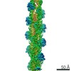
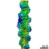
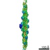
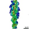
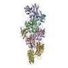
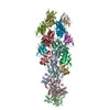
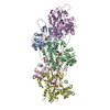
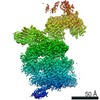
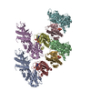
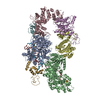
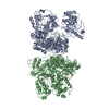
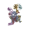
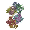
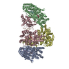
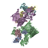
ジャーナル: Proc Natl Acad Sci U S A / 年: 2020 タイトル: Structures of cofilin-induced structural changes reveal local and asymmetric perturbations of actin filaments. 著者: Andrew R Huehn / Jeffrey P Bibeau / Anthony C Schramm / Wenxiang Cao / Enrique M De La Cruz / Charles V Sindelar / 要旨: Members of the cofilin/ADF family of proteins sever actin filaments, increasing the number of filament ends available for polymerization or depolymerization. Cofilin binds actin filaments with ...Members of the cofilin/ADF family of proteins sever actin filaments, increasing the number of filament ends available for polymerization or depolymerization. Cofilin binds actin filaments with positive cooperativity, forming clusters of contiguously bound cofilin along the filament lattice. Filament severing occurs preferentially at boundaries between bare and cofilin-decorated (cofilactin) segments and is biased at 1 side of a cluster. A molecular understanding of cooperative binding and filament severing has been impeded by a lack of structural data describing boundaries. Here, we apply methods for analyzing filament cryo-electron microscopy (cryo-EM) data at the single subunit level to directly investigate the structure of boundaries within partially decorated cofilactin filaments. Subnanometer resolution maps of isolated, bound cofilin molecules and an actin-cofilactin boundary indicate that cofilin-induced actin conformational changes are local and limited to subunits directly contacting bound cofilin. An isolated, bound cofilin compromises longitudinal filament contacts of 1 protofilament, consistent with a single cofilin having filament-severing activity. An individual, bound phosphomimetic (S3D) cofilin with weak severing activity adopts a unique binding mode that does not perturb actin structure. Cofilin clusters disrupt both protofilaments, consistent with a higher severing activity at boundaries compared to single cofilin. Comparison of these structures indicates that this disruption is substantially greater at pointed end sides of cofilactin clusters than at the barbed end. These structures, with the distribution of bound cofilin clusters, suggest that maximum binding cooperativity is achieved when 2 cofilins occupy adjacent sites. These results reveal the structural origins of cooperative cofilin binding and actin filament severing.
 ムービー
ムービー コントローラー


 データを開く
 基本情報
基本情報 要素
要素 キーワード
キーワード 機能・相同性情報
 Homo sapiens (ヒト)
 データ登録者
 米国, 2件
  引用
引用 構造の表示
構造の表示 ダウンロードとリンク
 その他のダウンロード
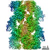

 PDBj
PDBj


 集合体
集合体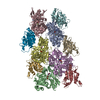


 分子量: 24.305 Da / 分子数: 5 / 由来タイプ: 合成 / 式: Mg
 分子量: 427.201 Da / 分子数: 5 / 由来タイプ: 合成 / 式: C10H15N5O10P2 / コメント: ADP, エネルギー貯蔵分子*YM
 試料調製
試料調製 電子顕微鏡撮影
 FIELD EMISSION GUN / 加速電圧: 300 kV / 照射モード: SPOT SCAN
 解析
解析