[English] 日本語
Yorodumi
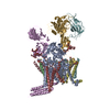
- PDB-6s7o: Cryo-EM structure of human oligosaccharyltransferase complex OST-A -

+
Open data


ID or keywords:

Loading...

-
Basic information

Entry
Database: PDB / ID: 6s7o
TitleCryo-EM structure of human oligosaccharyltransferase complex OST-A
Components
  • (Dolichyl-diphosphooligosaccharide--protein glycosyltransferase subunit ...) x 5
  • Dolichyl-diphosphooligosaccharide--protein glycosyltransferase 48 kDa subunit
  • Oligosaccharyltransferase complex subunit OSTCOligosaccharyltransferase
  • Transmembrane protein 258Transmembrane protein
KeywordsTRANSFERASE / N-glycosylation / Oligosaccharyltransferase / OSTA
Function / homology
Function and homology information


oligosaccharyltransferase complex binding / oligosaccharyltransferase I complex / oligosaccharyltransferase III complex / Asparagine N-linked glycosylation / membrane-bounded organelle / co-translational protein modification / oligosaccharyltransferase complex / dolichyl-diphosphooligosaccharide-protein glycotransferase / dolichyl-diphosphooligosaccharide-protein glycotransferase activity / protein N-linked glycosylation via asparagine ...oligosaccharyltransferase complex binding / oligosaccharyltransferase I complex / oligosaccharyltransferase III complex / Asparagine N-linked glycosylation / membrane-bounded organelle / co-translational protein modification / oligosaccharyltransferase complex / dolichyl-diphosphooligosaccharide-protein glycotransferase / dolichyl-diphosphooligosaccharide-protein glycotransferase activity / protein N-linked glycosylation via asparagine / protein N-linked glycosylation / epithelial cell apoptotic process / azurophil granule membrane / protein glycosylation / blastocyst development / Advanced glycosylation endproduct receptor signaling / SRP-dependent cotranslational protein targeting to membrane / enzyme activator activity / rough endoplasmic reticulum / response to endoplasmic reticulum stress / T cell activation / post-translational protein modification / response to cytokine / protein modification process / regulation of protein stability / melanosome / transferase activity / Maturation of spike protein / nuclear body / inflammatory response / intracellular membrane-bounded organelle / apoptotic process / Neutrophil degranulation / endoplasmic reticulum membrane / negative regulation of apoptotic process / endoplasmic reticulum / protein-containing complex / RNA binding / membrane / metal ion binding / plasma membrane / cytosol / cytoplasm
Similarity search - Function
DAD/Ost2 / Oligosaccharyltransferase complex subunit / DAD family / Oligosaccharyltransferase subunit 5 / Dolichyl-diphosphooligosaccharide--protein glycosyltransferase 48kDa subunit / Oligosaccharyltransferase complex subunit OSTC / Oligosaccharyltransferase 48 kDa subunit beta / Dolichyl-diphosphooligosaccharide--protein glycosyltransferase subunit Swp1 / Oligosaccharyltransferase subunit Ribophorin II / Oligosaccaryltransferase ...DAD/Ost2 / Oligosaccharyltransferase complex subunit / DAD family / Oligosaccharyltransferase subunit 5 / Dolichyl-diphosphooligosaccharide--protein glycosyltransferase 48kDa subunit / Oligosaccharyltransferase complex subunit OSTC / Oligosaccharyltransferase 48 kDa subunit beta / Dolichyl-diphosphooligosaccharide--protein glycosyltransferase subunit Swp1 / Oligosaccharyltransferase subunit Ribophorin II / Oligosaccaryltransferase / Oligosaccharyltransferase subunit ost4p superfamily / Oligosaccaryltransferase / Ribophorin I / Ribophorin I / Oligosaccharyl transferase complex, subunit OST3/OST6 / OST3 / OST6 family, transporter family / Oligosaccharyl transferase, STT3 subunit / Oligosaccharyl transferase STT3, N-terminal
Similarity search - Domain/homology
Chem-EGY / Chem-KZB / Chem-KZE / Dolichyl-diphosphooligosaccharide--protein glycosyltransferase 48 kDa subunit / Dolichyl-diphosphooligosaccharide--protein glycosyltransferase subunit 1 / Dolichyl-diphosphooligosaccharide--protein glycosyltransferase subunit 2 / Dolichyl-diphosphooligosaccharide--protein glycosyltransferase subunit 4 / Dolichyl-diphosphooligosaccharide--protein glycosyltransferase 48 kDa subunit / Dolichyl-diphosphooligosaccharide--protein glycosyltransferase subunit STT3A / Transmembrane protein 258 ...Chem-EGY / Chem-KZB / Chem-KZE / Dolichyl-diphosphooligosaccharide--protein glycosyltransferase 48 kDa subunit / Dolichyl-diphosphooligosaccharide--protein glycosyltransferase subunit 1 / Dolichyl-diphosphooligosaccharide--protein glycosyltransferase subunit 2 / Dolichyl-diphosphooligosaccharide--protein glycosyltransferase subunit 4 / Dolichyl-diphosphooligosaccharide--protein glycosyltransferase 48 kDa subunit / Dolichyl-diphosphooligosaccharide--protein glycosyltransferase subunit STT3A / Transmembrane protein 258 / Dolichyl-diphosphooligosaccharide--protein glycosyltransferase subunit DAD1 / Oligosaccharyltransferase complex subunit OSTC
Similarity search - Component
Biological speciesHomo sapiens (human)
MethodELECTRON MICROSCOPY / single particle reconstruction / cryo EM / Resolution: 3.5 Å
AuthorsRamirez, A.S. / Kowal, J. / Locher, K.P.
Funding support Switzerland, 1items
OrganizationGrant numberCountry
Swiss National Science FoundationCRSII3_147632 Switzerland
CitationJournal: Science / Year: 2019
Title: Cryo-electron microscopy structures of human oligosaccharyltransferase complexes OST-A and OST-B.
Authors: Ana S Ramírez / Julia Kowal / Kaspar P Locher /
Abstract: Oligosaccharyltransferase (OST) catalyzes the transfer of a high-mannose glycan onto secretory proteins in the endoplasmic reticulum. Mammals express two distinct OST complexes that act in a ...Oligosaccharyltransferase (OST) catalyzes the transfer of a high-mannose glycan onto secretory proteins in the endoplasmic reticulum. Mammals express two distinct OST complexes that act in a cotranslational (OST-A) or posttranslocational (OST-B) manner. Here, we present high-resolution cryo-electron microscopy structures of human OST-A and OST-B. Although they have similar overall architectures, structural differences in the catalytic subunits STT3A and STT3B facilitate contacts to distinct OST subunits, DC2 in OST-A and MAGT1 in OST-B. In OST-A, interactions with TMEM258 and STT3A allow ribophorin-I to form a four-helix bundle that can bind to a translating ribosome, whereas the equivalent region is disordered in OST-B. We observed an acceptor peptide and dolichylphosphate bound to STT3B, but only dolichylphosphate in STT3A, suggesting distinct affinities of the two OST complexes for protein substrates.
History
DepositionJul 5, 2019Deposition site: PDBE / Processing site: PDBE
Revision 1.0Dec 18, 2019Provider: repository / Type: Initial release
Revision 1.1Dec 25, 2019Group: Database references / Category: citation
Item: _citation.country / _citation.journal_abbrev ..._citation.country / _citation.journal_abbrev / _citation.journal_id_ASTM / _citation.journal_id_CSD / _citation.journal_id_ISSN / _citation.journal_volume / _citation.page_first / _citation.page_last / _citation.pdbx_database_id_DOI / _citation.pdbx_database_id_PubMed / _citation.title / _citation.year
Revision 2.0Jul 29, 2020Group: Advisory / Atomic model ...Advisory / Atomic model / Data collection / Derived calculations / Structure summary
Category: atom_site / chem_comp ...atom_site / chem_comp / entity / pdbx_branch_scheme / pdbx_chem_comp_identifier / pdbx_entity_branch / pdbx_entity_branch_descriptor / pdbx_entity_branch_link / pdbx_entity_branch_list / pdbx_entity_nonpoly / pdbx_nonpoly_scheme / pdbx_struct_assembly_gen / pdbx_unobs_or_zero_occ_atoms / pdbx_validate_close_contact / struct_asym / struct_conn / struct_site / struct_site_gen
Item: _atom_site.B_iso_or_equiv / _atom_site.Cartn_x ..._atom_site.B_iso_or_equiv / _atom_site.Cartn_x / _atom_site.Cartn_y / _atom_site.Cartn_z / _atom_site.auth_asym_id / _atom_site.auth_atom_id / _atom_site.auth_comp_id / _atom_site.auth_seq_id / _atom_site.label_asym_id / _atom_site.label_atom_id / _atom_site.label_comp_id / _atom_site.label_entity_id / _atom_site.type_symbol / _chem_comp.name / _chem_comp.type / _entity.formula_weight / _entity.pdbx_description / _entity.pdbx_number_of_molecules / _entity.type / _pdbx_struct_assembly_gen.asym_id_list / _pdbx_unobs_or_zero_occ_atoms.auth_asym_id / _pdbx_unobs_or_zero_occ_atoms.auth_seq_id / _pdbx_unobs_or_zero_occ_atoms.label_asym_id / _pdbx_validate_close_contact.auth_asym_id_1 / _pdbx_validate_close_contact.auth_asym_id_2 / _pdbx_validate_close_contact.auth_seq_id_1 / _pdbx_validate_close_contact.auth_seq_id_2 / _struct_conn.conn_type_id / _struct_conn.id / _struct_conn.pdbx_dist_value / _struct_conn.pdbx_leaving_atom_flag / _struct_conn.pdbx_role / _struct_conn.ptnr1_auth_asym_id / _struct_conn.ptnr1_auth_comp_id / _struct_conn.ptnr1_auth_seq_id / _struct_conn.ptnr1_label_asym_id / _struct_conn.ptnr1_label_atom_id / _struct_conn.ptnr1_label_comp_id / _struct_conn.ptnr1_label_seq_id / _struct_conn.ptnr2_auth_asym_id / _struct_conn.ptnr2_auth_comp_id / _struct_conn.ptnr2_auth_seq_id / _struct_conn.ptnr2_label_asym_id / _struct_conn.ptnr2_label_atom_id / _struct_conn.ptnr2_label_comp_id
Description: Carbohydrate remediation / Provider: repository / Type: Remediation

-
Structure visualization

Movie
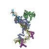
  • Deposited structure unit
  • Imaged by Jmol
  • Download
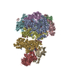
  • Superimposition on EM map
  • EMDB-10110
  • Imaged by UCSF Chimera
  • Download
Movie viewer
Structure viewerMolecule:
MolmilJmol/JSmol

Downloads & links

-
Assembly

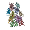
Deposited unit
A: Dolichyl-diphosphooligosaccharide--protein glycosyltransferase subunit STT3A
B: Dolichyl-diphosphooligosaccharide--protein glycosyltransferase subunit 4
C: Transmembrane protein 258
D: Dolichyl-diphosphooligosaccharide--protein glycosyltransferase subunit DAD1
E: Dolichyl-diphosphooligosaccharide--protein glycosyltransferase subunit 1
F: Dolichyl-diphosphooligosaccharide--protein glycosyltransferase subunit 2
G: Dolichyl-diphosphooligosaccharide--protein glycosyltransferase 48 kDa subunit
H: Oligosaccharyltransferase complex subunit OSTC
hetero molecules


Theoretical massNumber of molelcules
Total (without water)325,88630
Polymers311,9938
Non-polymers13,89322
Water0
1


  • Idetical with deposited unit
  • defined by author&software
  • Evidence: gel filtration
TypeNameSymmetry operationNumber
identity operation1_5551
Buried area41470 Å2
ΔGint-243 kcal/mol
Surface area100010 Å2
MethodPISA

-
Components

-
Dolichyl-diphosphooligosaccharide--protein glycosyltransferase subunit ... , 5 types, 5 molecules ABDEF

#1: Protein Dolichyl-diphosphooligosaccharide--protein glycosyltransferase subunit STT3A / STT3-A / B5 / Integral membrane protein 1 / Transmembrane protein TMC


Mass: 80607.320 Da / Num. of mol.: 1 / Source method: isolated from a natural source / Source: (natural) Homo sapiens (human)
References: UniProt: P46977, dolichyl-diphosphooligosaccharide-protein glycotransferase
#2: Protein/peptide Dolichyl-diphosphooligosaccharide--protein glycosyltransferase subunit 4


Mass: 4196.004 Da / Num. of mol.: 1 / Source method: isolated from a natural source / Source: (natural) Homo sapiens (human) / References: UniProt: P0C6T2
#4: Protein Dolichyl-diphosphooligosaccharide--protein glycosyltransferase subunit DAD1 / Oligosaccharyl transferase subunit DAD1 / Defender against cell death 1 / DAD-1


Mass: 12503.631 Da / Num. of mol.: 1 / Source method: isolated from a natural source / Source: (natural) Homo sapiens (human) / References: UniProt: P61803
#5: Protein Dolichyl-diphosphooligosaccharide--protein glycosyltransferase subunit 1 / Dolichyl-diphosphooligosaccharide--protein glycosyltransferase 67 kDa subunit / Ribophorin I / RPN- ...Dolichyl-diphosphooligosaccharide--protein glycosyltransferase 67 kDa subunit / Ribophorin I / RPN-I / Ribophorin-1


Mass: 68656.156 Da / Num. of mol.: 1 / Source method: isolated from a natural source / Source: (natural) Homo sapiens (human) / References: UniProt: P04843
#6: Protein Dolichyl-diphosphooligosaccharide--protein glycosyltransferase subunit 2 / Dolichyl-diphosphooligosaccharide--protein glycosyltransferase 63 kDa subunit / RIBIIR / Ribophorin ...Dolichyl-diphosphooligosaccharide--protein glycosyltransferase 63 kDa subunit / RIBIIR / Ribophorin II / RPN-II / Ribophorin-2


Mass: 69347.508 Da / Num. of mol.: 1 / Source method: isolated from a natural source / Source: (natural) Homo sapiens (human) / References: UniProt: P04844

-
Protein , 3 types, 3 molecules CGH

#3: Protein Transmembrane protein 258 / Transmembrane protein / Dolichyl-diphosphooligosaccharide--protein glycosyltransferase subunit TMEM258 / Oligosaccharyl ...Dolichyl-diphosphooligosaccharide--protein glycosyltransferase subunit TMEM258 / Oligosaccharyl transferase subunit TMEM258


Mass: 9083.804 Da / Num. of mol.: 1 / Source method: isolated from a natural source / Source: (natural) Homo sapiens (human) / References: UniProt: P61165
#7: Protein Dolichyl-diphosphooligosaccharide--protein glycosyltransferase 48 kDa subunit / Oligosaccharyl transferase 48 kDa subunit


Mass: 50754.438 Da / Num. of mol.: 1 / Source method: isolated from a natural source / Source: (natural) Homo sapiens (human) / References: UniProt: A0A024RAD5, UniProt: P39656*PLUS
#8: Protein Oligosaccharyltransferase complex subunit OSTC / Oligosaccharyltransferase / Hydrophobic protein HSF-28


Mass: 16844.215 Da / Num. of mol.: 1
Source method: isolated from a genetically manipulated source
Source: (gene. exp.) Homo sapiens (human) / Gene: OSTC, DC2, HDCMD45P, HSPC307 / Production host: Homo sapiens (human) / References: UniProt: Q9NRP0

-
Sugars , 3 types, 3 molecules

#9: Polysaccharide beta-D-mannopyranose-(1-4)-2-acetamido-2-deoxy-beta-D-glucopyranose-(1-4)-2-acetamido-2-deoxy-beta- ...beta-D-mannopyranose-(1-4)-2-acetamido-2-deoxy-beta-D-glucopyranose-(1-4)-2-acetamido-2-deoxy-beta-D-glucopyranose


Type: oligosaccharide / Mass: 586.542 Da / Num. of mol.: 1
Source method: isolated from a genetically manipulated source
DescriptorTypeProgram
DManpb1-4DGlcpNAcb1-4DGlcpNAcb1-ROHGlycam Condensed SequenceGMML 1.0
WURCS=2.0/2,3,2/[a2122h-1b_1-5_2*NCC/3=O][a1122h-1b_1-5]/1-1-2/a4-b1_b4-c1WURCSPDB2Glycan 1.1.0
[][D-1-deoxy-GlcpNAc]{[(4+1)][b-D-GlcpNAc]{[(4+1)][b-D-Manp]{}}}LINUCSPDB-CARE
#10: Polysaccharide alpha-D-mannopyranose-(1-2)-alpha-D-mannopyranose-(1-2)-alpha-D-mannopyranose-(1-3)-[alpha-D- ...alpha-D-mannopyranose-(1-2)-alpha-D-mannopyranose-(1-2)-alpha-D-mannopyranose-(1-3)-[alpha-D-mannopyranose-(1-6)-alpha-D-mannopyranose-(1-6)]beta-D-mannopyranose-(1-4)-2-acetamido-2-deoxy-beta-D-glucopyranose-(1-4)-2-acetamido-2-deoxy-beta-D-glucopyranose


Type: oligosaccharide / Mass: 1397.245 Da / Num. of mol.: 1
Source method: isolated from a genetically manipulated source
DescriptorTypeProgram
DManpa1-2DManpa1-2DManpa1-3[DManpa1-6DManpa1-6]DManpb1-4DGlcpNAcb1-4DGlcpNAcb1-Glycam Condensed SequenceGMML 1.0
WURCS=2.0/3,8,7/[a2122h-1b_1-5_2*NCC/3=O][a1122h-1b_1-5][a1122h-1a_1-5]/1-1-2-3-3-3-3-3/a4-b1_b4-c1_c3-d1_c6-g1_d2-e1_e2-f1_g6-h1WURCSPDB2Glycan 1.1.0
[]{[(4+1)][b-D-GlcpNAc]{[(4+1)][b-D-GlcpNAc]{[(4+1)][b-D-Manp]{[(3+1)][a-D-Manp]{[(2+1)][a-D-Manp]{[(2+1)][a-D-Manp]{}}}[(6+1)][a-D-Manp]{[(6+1)][a-D-Manp]{}}}}}}LINUCSPDB-CARE
#11: Polysaccharide alpha-D-mannopyranose-(1-2)-alpha-D-mannopyranose-(1-3)-[alpha-D-mannopyranose-(1-3)-[alpha-D- ...alpha-D-mannopyranose-(1-2)-alpha-D-mannopyranose-(1-3)-[alpha-D-mannopyranose-(1-3)-[alpha-D-mannopyranose-(1-6)]alpha-D-mannopyranose-(1-6)]beta-D-mannopyranose-(1-4)-2-acetamido-2-deoxy-beta-D-glucopyranose-(1-4)-2-acetamido-2-deoxy-beta-D-glucopyranose


Type: oligosaccharide / Mass: 1397.245 Da / Num. of mol.: 1
Source method: isolated from a genetically manipulated source
DescriptorTypeProgram
DManpa1-2DManpa1-3[DManpa1-3[DManpa1-6]DManpa1-6]DManpb1-4DGlcpNAcb1-4DGlcpNAcb1-Glycam Condensed SequenceGMML 1.0
WURCS=2.0/3,8,7/[a2122h-1b_1-5_2*NCC/3=O][a1122h-1b_1-5][a1122h-1a_1-5]/1-1-2-3-3-3-3-3/a4-b1_b4-c1_c3-d1_c6-f1_d2-e1_f3-g1_f6-h1WURCSPDB2Glycan 1.1.0
[]{[(4+1)][b-D-GlcpNAc]{[(4+1)][b-D-GlcpNAc]{[(4+1)][b-D-Manp]{[(3+1)][a-D-Manp]{[(2+1)][a-D-Manp]{}}[(6+1)][a-D-Manp]{[(3+1)][a-D-Manp]{}[(6+1)][a-D-Manp]{}}}}}}LINUCSPDB-CARE

-
Non-polymers , 4 types, 19 molecules
#12: Chemical
ChemComp-KZB / (2~{S},3~{R},4~{R},5~{S},6~{S})-2-(hydroxymethyl)-6-[(1~{S},2~{R},3~{R},4~{R},5'~{S},6~{S},7~{R},8~{S},9~{R},12~{R},13~{R},15~{S},16~{S},18~{R})-5',7,9,13-tetramethyl-3,15-bis(oxidanyl)spiro[5-oxapentacyclo[10.8.0.0^{2,9}.0^{4,8}.0^{13,18}]icosane-6,2'-oxane]-16-yl]oxy-oxane-3,4,5-triol


Mass: 610.776 Da / Num. of mol.: 9
Source method: isolated from a genetically manipulated source
Formula: C33H54O10
#13: Chemical
ChemComp-EGY / (4R,7R)-4-hydroxy-N,N,N-trimethyl-4,9-dioxo-7-[(undecanoyloxy)methyl]-3,5,8-trioxa-4lambda~5~-phosphadocosan-1-aminium


Mass: 636.861 Da / Num. of mol.: 7
Source method: isolated from a genetically manipulated source
Formula: C33H67NO8P / Comment: phospholipid*YM
#14: Chemical ChemComp-MG / MAGNESIUM ION


Mass: 24.305 Da / Num. of mol.: 2
Source method: isolated from a genetically manipulated source
Formula: Mg
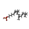
#15: Chemical ChemComp-KZE / [(3~{R},6~{Z},10~{Z},14~{Z},18~{Z})-3,7,11,15,19,23-hexamethyltetracosa-6,10,14,18,22-pentaenyl] dihydrogen phosphate


Mass: 508.713 Da / Num. of mol.: 1
Source method: isolated from a genetically manipulated source
Formula: C30H53O4P

-
Details

Has ligand of interestN

-
Experimental details

-
Experiment

ExperimentMethod: ELECTRON MICROSCOPY
EM experimentAggregation state: PARTICLE / 3D reconstruction method: single particle reconstruction

-
Sample preparation

Component
IDNameTypeEntity IDParent-IDSource
1Human oligosaccharyltransferase complex OST-ACOMPLEX#1-#80MULTIPLE SOURCES
2Human oligosaccharyltransferase complexOligosaccharyltransferaseCOMPLEX#1-#71NATURAL
3Oligosaccharyltransferase complex subunit OSTCOligosaccharyltransferaseCOMPLEX#81RECOMBINANT
Molecular weightExperimental value: NO
Source (natural)
IDEntity assembly-IDOrganismNcbi tax-ID
22Homo sapiens (human)9606
33Homo sapiens (human)9606
Source (recombinant)Organism: Homo sapiens (human)
Buffer solutionpH: 7.5
SpecimenEmbedding applied: NO / Shadowing applied: NO / Staining applied: NO / Vitrification applied: YES
VitrificationCryogen name: ETHANE-PROPANE

-
Electron microscopy imaging

Experimental equipment
Model: Titan Krios / Image courtesy: FEI Company
MicroscopyModel: FEI TITAN KRIOS
Electron gunElectron source: FIELD EMISSION GUN / Accelerating voltage: 300 kV / Illumination mode: FLOOD BEAM
Electron lensMode: BRIGHT FIELDBright-field microscopy / Cs: 2.7 mm / Alignment procedure: COMA FREE
Specimen holderCryogen: NITROGEN / Specimen holder model: FEI TITAN KRIOS AUTOGRID HOLDER
Image recordingAverage exposure time: 8 sec. / Electron dose: 68 e/Å2 / Detector mode: COUNTING / Film or detector model: GATAN K2 SUMMIT (4k x 4k) / Num. of grids imaged: 3 / Num. of real images: 6035
EM imaging opticsEnergyfilter name: GIF Quantum LS / Energyfilter slit width: 20 eV
Image scansMovie frames/image: 40

-
Processing

EM software
IDNameVersionCategory
2SerialEMimage acquisition
4GctfCTF correction
10RELION3initial Euler assignment
11RELION3final Euler assignment
12RELION3classification
13RELION33D reconstruction
CTF correctionType: PHASE FLIPPING AND AMPLITUDE CORRECTION
Particle selectionNum. of particles selected: 424817
SymmetryPoint symmetry: C1 (asymmetric)
3D reconstructionResolution: 3.5 Å / Resolution method: FSC 0.143 CUT-OFF / Num. of particles: 156950 / Symmetry type: POINT

+
About Yorodumi

-
News

-
Feb 9, 2022. New format data for meta-information of EMDB entries

New format data for meta-information of EMDB entries

  • Version 3 of the EMDB header file is now the official format.
  • The previous official version 1.9 will be removed from the archive.

Related info.:EMDB header

External links:wwPDB to switch to version 3 of the EMDB data model

-
Aug 12, 2020. Covid-19 info

Covid-19 info

URL: https://pdbj.org/emnavi/covid19.php

New page: Covid-19 featured information page in EM Navigator.

Related info.:Covid-19 info / Mar 5, 2020. Novel coronavirus structure data

+
Mar 5, 2020. Novel coronavirus structure data

Novel coronavirus structure data

Related info.:Yorodumi Speices / Aug 12, 2020. Covid-19 info

External links:COVID-19 featured content - PDBj / Molecule of the Month (242):Coronavirus Proteases

+
Jan 31, 2019. EMDB accession codes are about to change! (news from PDBe EMDB page)

EMDB accession codes are about to change! (news from PDBe EMDB page)

  • The allocation of 4 digits for EMDB accession codes will soon come to an end. Whilst these codes will remain in use, new EMDB accession codes will include an additional digit and will expand incrementally as the available range of codes is exhausted. The current 4-digit format prefixed with “EMD-” (i.e. EMD-XXXX) will advance to a 5-digit format (i.e. EMD-XXXXX), and so on. It is currently estimated that the 4-digit codes will be depleted around Spring 2019, at which point the 5-digit format will come into force.
  • The EM Navigator/Yorodumi systems omit the EMD- prefix.

Related info.:Q: What is EMD? / ID/Accession-code notation in Yorodumi/EM Navigator

External links:EMDB Accession Codes are Changing Soon! / Contact to PDBj

+
Jul 12, 2017. Major update of PDB

Major update of PDB

  • wwPDB released updated PDB data conforming to the new PDBx/mmCIF dictionary.
  • This is a major update changing the version number from 4 to 5, and with Remediation, in which all the entries are updated.
  • In this update, many items about electron microscopy experimental information are reorganized (e.g. em_software).
  • Now, EM Navigator and Yorodumi are based on the updated data.

External links:wwPDB Remediation / Enriched Model Files Conforming to OneDep Data Standards Now Available in the PDB FTP Archive

-
Yorodumi

Thousand views of thousand structures

  • Yorodumi is a browser for structure data from EMDB, PDB, SASBDB, etc.
  • This page is also the successor to EM Navigator detail page, and also detail information page/front-end page for Omokage search.
  • The word "yorodu" (or yorozu) is an old Japanese word meaning "ten thousand". "mi" (miru) is to see.

Related info.:EMDB / PDB / SASBDB / Comparison of 3 databanks / Yorodumi Search / Aug 31, 2016. New EM Navigator & Yorodumi / Yorodumi Papers / Jmol/JSmol / Function and homology information / Changes in new EM Navigator and Yorodumi

Read more