 Movie
Movie Controller

[English] 日本語
 Yorodumi
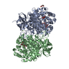
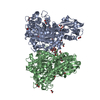
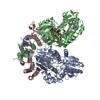
- PDB-6ts8: Chaetomium thermophilum UDP-Glucose Glucosyl Transferase (UGGT) d... -
+ Open data

- Basic information
| Entry | Database: PDB / ID: 6ts8 | ||||||||||||
|---|---|---|---|---|---|---|---|---|---|---|---|---|---|
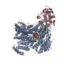
| Title | Chaetomium thermophilum UDP-Glucose Glucosyl Transferase (UGGT) double cysteine mutant G177C/A786C. | ||||||||||||
 Components Components | UDP-glucose-glycoprotein glucosyltransferase-like protein | ||||||||||||
 Keywords Keywords | PROTEIN BINDING / Endoplasmic Reticulum / Glycoprotein Folding / ERQC / UGGT | ||||||||||||
| Function / homology |  Function and homology information Function and homology informationUDP-glucose:glycoprotein glucosyltransferase activity / glycoprotein biosynthetic process / ERAD pathway / : / endoplasmic reticulum lumen / nucleotide binding / metal ion binding Similarity search - Function | ||||||||||||
| Biological species |  Chaetomium thermophilum (fungus) Chaetomium thermophilum (fungus) | ||||||||||||
| Method |  X-RAY DIFFRACTION / SYNCHROTRON / MOLECULAR REPLACEMENT / Resolution: 4.6 Å X-RAY DIFFRACTION / SYNCHROTRON / MOLECULAR REPLACEMENT / Resolution: 4.6 Å | ||||||||||||
 Authors Authors | Roversi, P. / Zitzmann, N. / Ibba, R. / Hensen, M. / Chandran, A. | ||||||||||||
| Funding support |  United Kingdom, 3items United Kingdom, 3items
| ||||||||||||
 Citation Citation | Journal: Structure / Year: 2021 Title: Clamping, bending, and twisting inter-domain motions in the misfold-recognizing portion of UDP-glucose: Glycoprotein glucosyltransferase. Authors: Modenutti, C.P. / Blanco Capurro, J.I. / Ibba, R. / Alonzi, D.S. / Song, M.N. / Vasiljevic, S. / Kumar, A. / Chandran, A.V. / Tax, G. / Marti, L. / Hill, J.C. / Lia, A. / Hensen, M. / ...Authors: Modenutti, C.P. / Blanco Capurro, J.I. / Ibba, R. / Alonzi, D.S. / Song, M.N. / Vasiljevic, S. / Kumar, A. / Chandran, A.V. / Tax, G. / Marti, L. / Hill, J.C. / Lia, A. / Hensen, M. / Waksman, T. / Rushton, J. / Rubichi, S. / Santino, A. / Marti, M.A. / Zitzmann, N. / Roversi, P. | ||||||||||||
| History |
|
- Structure visualization
| Structure viewer | Molecule: MolmilJmol/JSmol |
|---|
- Downloads & links
-Download
| PDBx/mmCIF format | 6ts8.cif.gz | 1010.9 KB | Display | PDBx/mmCIF format |
|---|---|---|---|---|
| PDB format | pdb6ts8.ent.gz | 838.1 KB | Display | PDB format |
| PDBx/mmJSON format | 6ts8.json.gz | Tree view | PDBx/mmJSON format | |
| Others |  Other downloads Other downloads |
-Validation report
| Arichive directory | https://data.pdbj.org/pub/pdb/validation_reports/ts/6ts8ftp://data.pdbj.org/pub/pdb/validation_reports/ts/6ts8 | HTTPS FTP |
|---|
-Related structure data
| Related structure data | 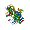 6trfC  6trtC 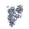 6ts2C 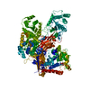 5nv4S S: Starting model for refinement C: citing same article ( |
|---|---|
| Similar structure data |


-Links
 PDBj
PDBj


- Assembly
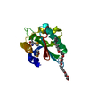
| Deposited unit | 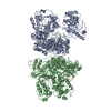
| ||||||||||||||||||
|---|---|---|---|---|---|---|---|---|---|---|---|---|---|---|---|---|---|---|---|
| 1 | 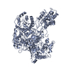
| ||||||||||||||||||
| 2 | 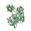
| ||||||||||||||||||
| Unit cell |
| ||||||||||||||||||
| Noncrystallographic symmetry (NCS) | NCS domain:
NCS domain segments: Component-ID: _ / Ens-ID: 1 / Beg auth comp-ID: SER / Beg label comp-ID: SER / End auth comp-ID: ILE / End label comp-ID: ILE / Refine code: _ / Auth seq-ID: 28 - 1409 / Label seq-ID: 1 - 1382
|
-Components
| #1: Protein | Mass: 156354.812 Da / Num. of mol.: 2 Source method: isolated from a genetically manipulated source Details: Chaetomium thermophilum UDP-Glucose Glucosyl Transferase (UGGT) double cysteine mutant G177C/A786C. Source: (gene. exp.) Chaetomium thermophilum (strain DSM 1495 / CBS 144.50 / IMI 039719) (fungus)Strain: DSM 1495 / CBS 144.50 / IMI 039719 / Gene: CTHT_0048990 / Plasmid: pHLsec / Cell (production host): HEK293F / Production host:  Homo sapiens (human) / References: UniProt: G0SB58 Homo sapiens (human) / References: UniProt: G0SB58Has ligand of interest | N | Has protein modification | Y | |
|---|
-Experimental details
-Experiment
| Experiment | Method: X-RAY DIFFRACTION / Number of used crystals: 1 |
|---|
- Sample preparation
| Crystal | Density Matthews: 2.47 Å3/Da / Density % sol: 50.17 % |
|---|---|
| Crystal grow | Temperature: 291 K / Method: vapor diffusion, sitting drop / pH: 5.8 Details: 16.54% w/v PEG 4,000, 0.03 M citric acid pH 5.3, 0.07 M citric Acid pH 6.0, 12.75% v/v isopropanol |
-Data collection
| Diffraction | Mean temperature: 80 K / Serial crystal experiment: N |
|---|---|
| Diffraction source | Source: SYNCHROTRON / Site: Diamond / Beamline: I04 / Wavelength: 0.97949 Å |
| Detector | Type: DECTRIS PILATUS 6M / Detector: PIXEL / Date: Oct 8, 2018 |
| Radiation | Protocol: SINGLE WAVELENGTH / Monochromatic (M) / Laue (L): M / Scattering type: x-ray |
| Radiation wavelength | Wavelength: 0.97949 Å / Relative weight: 1 |
| Reflection | Resolution: 4.59→139.05 Å / Num. obs: 22051 / % possible obs: 87.8 % / Redundancy: 4.3 % / CC1/2: 0.982 / Rmerge(I) obs: 0.278 / Rpim(I) all: 0.147 / Rrim(I) all: 0.316 / Net I/σ(I): 4.6 |
| Reflection shell | Resolution: 4.717→5.576 Å / Redundancy: 6.8 % / Rmerge(I) obs: 1.255 / Mean I/σ(I) obs: 1.5 / Num. unique obs: 1744 / CC1/2: 0.589 / Rpim(I) all: 0.52 / Rrim(I) all: 1.338 / % possible all: 69.3 |
- Processing
| Software |
| ||||||||||||||||||||||||||||||||||||||||||||||||||||||||||||||||||||||||||||||||||||||||||||||||||||||||||||||||||||||||||||||||||||||||||||||||||||||||||||||||||||||||||||||||||||||
|---|---|---|---|---|---|---|---|---|---|---|---|---|---|---|---|---|---|---|---|---|---|---|---|---|---|---|---|---|---|---|---|---|---|---|---|---|---|---|---|---|---|---|---|---|---|---|---|---|---|---|---|---|---|---|---|---|---|---|---|---|---|---|---|---|---|---|---|---|---|---|---|---|---|---|---|---|---|---|---|---|---|---|---|---|---|---|---|---|---|---|---|---|---|---|---|---|---|---|---|---|---|---|---|---|---|---|---|---|---|---|---|---|---|---|---|---|---|---|---|---|---|---|---|---|---|---|---|---|---|---|---|---|---|---|---|---|---|---|---|---|---|---|---|---|---|---|---|---|---|---|---|---|---|---|---|---|---|---|---|---|---|---|---|---|---|---|---|---|---|---|---|---|---|---|---|---|---|---|---|---|---|---|---|
| Refinement | Method to determine structure: MOLECULAR REPLACEMENT Starting model: 5nv4 Resolution: 4.6→139.04 Å / Cor.coef. Fo:Fc: 0.892 / Cor.coef. Fo:Fc free: 0.887 / SU B: 258.499 / SU ML: 1.351 / Cross valid method: THROUGHOUT / ESU R Free: 2.993 / Stereochemistry target values: MAXIMUM LIKELIHOOD / Details: HYDROGENS HAVE BEEN ADDED IN THE RIDING POSITIONS
| ||||||||||||||||||||||||||||||||||||||||||||||||||||||||||||||||||||||||||||||||||||||||||||||||||||||||||||||||||||||||||||||||||||||||||||||||||||||||||||||||||||||||||||||||||||||
| Solvent computation | Ion probe radii: 0.8 Å / Shrinkage radii: 0.8 Å / VDW probe radii: 1.2 Å / Solvent model: MASK | ||||||||||||||||||||||||||||||||||||||||||||||||||||||||||||||||||||||||||||||||||||||||||||||||||||||||||||||||||||||||||||||||||||||||||||||||||||||||||||||||||||||||||||||||||||||
| Displacement parameters | Biso mean: 160.355 Å2
| ||||||||||||||||||||||||||||||||||||||||||||||||||||||||||||||||||||||||||||||||||||||||||||||||||||||||||||||||||||||||||||||||||||||||||||||||||||||||||||||||||||||||||||||||||||||
| Refinement step | Cycle: 1 / Resolution: 4.6→139.04 Å
| ||||||||||||||||||||||||||||||||||||||||||||||||||||||||||||||||||||||||||||||||||||||||||||||||||||||||||||||||||||||||||||||||||||||||||||||||||||||||||||||||||||||||||||||||||||||
| Refine LS restraints |
|